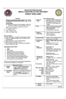
Histopathologic and Cytologic Techniques Lecture Notes PDF

| Title | Histopathologic and Cytologic Techniques Lecture Notes |
|---|---|
| Author | Anonymous User |
| Course | Ethics |
| Institution | Davao Doctors College |
| Pages | 45 |
| File Size | 2.8 MB |
| File Type | |
| Total Downloads | 192 |
| Total Views | 702 |
Summary
Download Histopathologic and Cytologic Techniques Lecture Notes PDF
Description
DAVAO DOCTORS COLLEGE MEDICAL LABORATORY SCIENCE DEPARTMENT
STUDENT NOTES: GPHCT REVIEW OF BASIC HISTOLOGY
INTRODUCTION TO GENERAL PATHOLOGY
Histology
Study of tissues and their structure.
Pathology (pathos: suffering; logos: study)
Germ Layers A. Ectoderm B. Mesoderm C. Endoderm
Group of cells that form during embryonic development. The layers will eventually differentiate into various tissues and organs.
study of diseases at cellular, tissue and organ level bridge between mediZcine and science; it is the scientific foundation of medicine
Types of Tissues A. Epithelial
Covering epithelia: Lines surfaces Glandular epithelia: secrete substances
B. Connective
Most abundant; Binds, protects, supports
C. Muscular
Highly specialized; Movement
D. Nervous
Sensation, integration, response
Importance of Histology in General Pathology
Disease processes affect tissues in distinctive ways, which depend on the type of tissue, and the disease itself.
These processes may cause them to die, to change their shape, to divide, to move or to invade other tissues. Any of these changes also affect the anatomy of the tissue.
Understanding the changes that are characteristic of a disease requires a detailed knowledge of the normal histology of cells and tissues, and the range of normality.
Knowing the type of tissue and their composition is important in the selection of the appropriate histopathologic technique and stain to be used.
These changes within cells and tissues can be visualized using histopathologic techniques.
-
Divisions of Pathology A. Gross Pathology
Macroscopic examination of tissues and organs
B. Microscopic Pathology i. Anatomic Pathology
Surgical Biopsy (living), Autopsy (dead) Histopathology ii. Clinical Pathology • Hematology • Microbiology • Clinical Chemistry • Immunology/Serology • Clinical Microscopy • Parasitology Rudolf Virchow – father of modern pathology • •
Disease -
Any change from a state of health as a result of certain forms of stimuli and stress, which leads to impaired physiological functioning
Four Aspects of a Disease Process Cause or origin of the disease; might be Etiology genetic factors or acquired factors Pathogenesis Mechanisms of the development of the disease Sequence of events from initial stimulus to ultimate expression of the disease How etiologic factors trigger cellular & molecular changes in a disease Morphologic & Molecular Changes Clinical Manifestations Signs Symptoms
Structural, biochemical and molecular alterations induced in the cells and organs of the body as a result of the disease Functional consequence of the changes Effects that can be observed by others Effects apparent only to the patient
Almost all forms of disease start with alterations in cells. Therefore, it is important to study the causes, mechanisms and morphologic and biochemical correlates of cell injury. JALN2020
Stages of the Cellular Response to Stress and Injurious Stimuli
CELLULAR ADAPTATION -
Changes made by a cell in response to stress or stimuli May be physiologic or pathologic Forms of Adaptation 1. Hypertrophy Increased Cell Size Increased Organ Size Due to increased protein synthesis Most common stimulus: Increased Workload Examples: bulging muscles of bodybuilders, estrogen-induced enlargement of uterus during pregnancy Remember: *body builders win trophy* Hyperplasia
2.
Increased Cell Number Increased Organ Mass Due to proliferative actions of growth factor, and/or stem cells 1.
Normal cells have defined structure and can perform limited functions based on their specialization, metabolism, and availability of metabolic substrates. They can handle physiologic demands through homeostasis.
Physiologic: Hormonal hyperplasia - Breast during puberty or pregnancy
Compensatory hyperplasia - Liver cells regeneration
Pathologic: Homeostasis - act of maintaining a steady state 2.
When there is a slightly severe stress, or some pathologic stimuli, cells undergo adaptation in order to survive and continue to function. Adaptation - reversible structural and functional response of cells to stress and stimuli
3.
Excess Hormonal Stimulation Increased Estrogen Endometrial hyperplasia abnormal menstrual bleeding ■ Excess Growth Hormone Stimulation Papillomavirus mucosal lesions ■
But if the limits of adaptive response are exceeded, or when cells are exposed to injurious stimuli (agents or stress), or deprived of essential nutrients, cell injury occurs. a. If the stimulus is mild and transient, the injury is reversible. The cell may go back to its normal state. b. If it is severe and progressive, the injury is irreversible. Cells that undergo irreversible injury will ultimately suffer cell death, which may be pathological or physiological.
Other types of stress can induce responses other than cellular adaptation, injury and death. The responses are the following: Autophagy (self-eating) - starved cells eat its own components during nutrient deprivation B. Intracellular accumulation of substances (such as proteins, lipids, hyaline, glycogen, pigments) C. Pathologic calcification – abnormal tissue deposition of calcum salts D. Cellular aging – progressive decline in the life span and functional capacity of cells
3.
Atrophy Decreased Cell Size & Number Reduce tissue/organ size Due to decreased protein synthesis, and increased protein degradation
Physiologic: as in puberty when the thymus and the lymphoid organs decrease in size e.g. Atrophy of uterus after pregnancy Pathologic (Types): 1. Atrophy of disuse - decreased workload, thus diminished function of organ e.g. skeletal muscle atrophy due to bedrest 2.
Vascular - Diminished blood supply (ischemia) e.g. brain atrophy during atherosclerosis
3.
Starvation - inadequate nutrition of cell e.g. muscle wasting (or cachexia) due to use of skeletal muscle as source of energy during protein malnutrition
4.
Loss of endocrine stimulation - due to decrease of regulating hormones e.g. breast atrophy after menopause due to loss of estrogen stimulation
5.
Pressure - as in growth of tumors, atrophy occurs when tumors suppress the blood supply or by directly putting pressure to surrounding healthy cells
6.
Exhaustion - due to increase in metabolism resulting to increase of metabolites and loss of the actual cell space
A.
JALN2020
Metaplasia Change in one cell type to another Due to reprogramming of existing stem cells in normal tissue, or of undifferentiated mesenchymal cells in order to withstand adverse environment
4.
Example: > Habitual cigarette smokers - ciliated columnar cells of trachea and bronchi are replaced by stratified squamous cells > Barrett esophagus - squamous cells of esophagus are replaced by intestinal-like columnar cells in response to refluxed acid
APOPTOSIS Induced by a tightly regulated suicide program in which cells destined to die activate enzymes that degrade the cells' own proteins and nuclear DNA Presence of cleaved, active caspases (cysteine proteases that cleave aspartic acid residue) is a marker for cells undergoing apoptosis Cells break up into apoptotic bodies, which are tasty targets for phagocytes Reasons for Apoptosis in Following Conditions: Physiologic Pathologic
CELLULAR INJURY -
Alteration in cell structure or function due to stress or pathologic stimuli
Eliminates cells that are no longer needed, or those that have served their purposes Eliminates cells that are injured beyond repair without eliciting host reaction
NECROSIS - Pathologic cell death Necrotic - tissue or organs with large numbers of dead cells
Causes Hypoxia Physical Agents Chemical Agents and Drugs Infectious Agents
Immunologic Reactions Genetic Abnormalities Nutritional Imbalances
Morphological Alterations in Cell Injury Generalized swelling of cell and organelles - first manifestation Blebbing of plasma membrane Detachment of ribosome from ER Clumping of nuclear chromatin
-
CELL DEATH Occurs after irreversible injury
Difference between the Two Principal Pathways of Cell Death
Cell Size Nucleus
Plasma Membrane Cellular contents Adjacent Inflammation
Physiologic or Pathologic?
Types of Necrosis According to Morphology 1. Coagulative Tissue is firm because architecture of dead tissue is preserved Eosinophilic due to denaturation of proteins AND enzymes Occurs on affected tissue when vessel is obstructed, except brain Infarct - localized area of coagulative necrosis 2. Liquefactive Tissue becomes liquid viscous mass due to digestion of dead cells Occurs during microbial infection Creamy yellow because of pus Affects CNS 3. Gangrenous Due to ischemia and superimposed bacterial infection Combination of coagulation and liquefaction necrosis Sterile necrosis i. Dry Gangrene Arterial occlusion, sharp demarcation line, less foul odor
Apoptosis
Necrosis
Reduced Fragmentation into nucleosome-size fragments Intact
Enlarged Pyknosis (clumping) Karyorrhexis (fragmentation) Karyolysis (dissolution) Disrupted
Intact
Enzymatic digestion; may leak out of cell Frequent (due to leakage of cellular contents)
5. Fat
Pathologic
6. Fibrinoid
ii. Wet Gangrene 4. Caseous
No (because phagocytes rapidly devour the cells) Physiologic Death by destiny
Death by disease
Nonsterile necrosis (due to bacterial infection) Vein occlusion, no sharp demarcation line, foul odor Means ‘cheese-like’ Friable white appearance of necrotic area Seen in tuberculosis, granuloma Fat destruction due to pancreatic lipase Fatty Acids + Calcium = Chalky-white areas (fat saponification) Seen in acute pancreatitis Seen in immune reactions when antigen- antibody complexes are deposited in walls of arteries Immune complex + fibrin = fibrinoid (bright pink and amorphous appearance in H&E staining)
JALN2020
INFLAMMATION -
A protective universal response to tissue damage (mechanical trauma, tissue necrosis, infection) May be beneficial or harmful Functions: 1. contain damage & isolate injury 2. destroy cause of injury (microorganism/toxins) 3. destroy resulting necrotic cells and tissues 4. prepare tissue for healing & repair
Classes of Inflammation 1.
i. Acute
2.
Harmful effects: 1. Digestion of normal tissues 2. Swelling 3. Inappropriate inflammatory response Cardinal Signs: 1. Rubor – redness 2. Calor – heat 3. Tumor – swelling 4. Dolor – pain 5. Functio laesa – loss of function
Vascular Reaction i. Vasoconstriction ii. Vasodilation
iii. Endothelial Activation 2.
Cellular Response i. Neutrophil Activation
WBCs enter site of injury Kill organism, mop debris Release chemokines (substances that attract other immune substances to site of inflammation)
i. Serous
Out-pouring of relatively protein-poor fluid; common in cavities
ii. Fibrinous
More severe injury more vascular permeability more protein leaking (such as fibrinogen which is the precursor of fibrin) Increased blood flow to mucosal vessels, enlargement of secretory vessels discharge of mucus and epithelial debris Disruption of blood vessel wall leakage of large number of RBCs
iv. Hemorrhagic v. Suppurative / Purulent
Occurs first and lasts only for seconds Increased diameter of blood vessels Increased blood flow to area Erythema (heat and redness on site of infection) Increased vascular permeability Edema (extravasation of liquid portion of blood)
Sudden onset; usually mild and selflimiting; Polymorphonuclear (PMNs) cells ii. Chronic Involves persistence of injurious agent; often severe and progressive; Mononuclear cells According to Character of Exudate
iii. Catarrhal
Components of Inflammation 1.
According to Duration
Mainly due to bacterial infection or secondary condition Collection of large amount of Pus Composed of : Neutrophil Necrotic cells Edema fluid Abscess = collection of pus
3.
According to Location i. Localized
One site; not widespread
ii. Generalized / Systemic
Whole organ; area of tissue; region of tissue
Sequential Steps of a Typical Inflammatory Reaction 1. The offending agent is recognized by host cells and molecules. 2. WBCs and plasma proteins are recruited from the circulation (Intravascular), and go towards the area where the offending agent is located. 3. WBCs and proteins are activated and work together to destroy and eliminate the offending substance. 4. Reaction is controlled and terminated. 5. Damaged tissue is repaired Note: If inflammatory reaction is not controlled, adverse tissue effects may occur, such as abscess formation and chronic inflammation.
JALN2020
ABNORMALITIES IN CELL GROWTH I.
Retrogressive Changes - organs are smaller than the normal A. Developmental Defects 1. Aplasia - incomplete development of the organ 2. Hypoplasia - failure of an organ to develop fully 3. Agenesia - complete non-appearance of an organ 4. Atresia - failure of an organ to form an opening
Classification of Tumor Death
Differentiation Rate of Growth
B.
II.
III.
Atrophy - acquired decrease of the size of a normally developed organ
Progressive Changes - organs become larger than normal A. Hypertrophy - increase of cell size 1. True - due to increase work load or endocrine stimulation a. Compensatory - true for paired organs, where one is excised and the other incresases in size to “take responsibility’ for its pair 2. False - due to ECF buildup and CT proliferation B. Hyperplasia - increase in cell population 1. Physiologic, as in pregnancy 2. Pathologic, as in typhoid fever affecting lymphoid follicles Degenerative Changes – changes in the adult form of cell A. Metaplasia - replacement of one cell type of cells to another type in the same site (reversible) B. Dysplasia – means “disordered growth”; development of abnormal cell types within a tissue (reversible) C. Anaplasia – lack of differentiation of cells (irreversible) D. Neoplasia – means “new growth”; uncontrolled proliferation of cells with no purpose; due to carcinogens, or DNA alteration (irreversible)
Tumor/Neoplasms – mass of neoplastic cells
General Characteristics of Tumors 1. May resemble and function like a normal cell 2. Autonomous; non-responsive to normal growth factors 3. Parasitic nature; competes with cells for metabolic needs Parts of Tumor 1. Parenchyma – actively dividing cells 2. Stroma –connective tissue framework and lymphatic and vascular channels
Metastasis (spread of tumor to other sites)
Benign Usually does not cause death except for infants and brain tumors Well-differentiated Usually progressive and slow; may come to a standstill or regress Absent
General Tumor Nomenclature Benign Origin Epithelial Tissue Connective/ -oma Mesenchyme Tissue Example: Epithelial lining of gland Lymph vessels
Malignant Usually causes death
Some lack of differentiation Erratic; may be slow to rapid
Frequent
Malignant -carcinoma -sarcoma
Benign Adenoma
Malignant Adenocarcinoma
Lymphangioma
Lymphangiosarcoma
Grading of Tumors -
Attempts to establish an estimate of the level of malignancy of a tumor Based on cytologic differentiation of tumor cells and number of mitoses Different from Staging, as staging accounts for size, extent of spread to lymph nodes, and presence of metastasis Well-differentiated: tumor cells resemble normal cells Undifferentiated: tumor cells do not resemble normal cells
Broder’s Grading of Tumors Grade Differentiated Cells I 100% - 75% II 75% - 50% III 50% - 25% IV 25% - 0%
Undifferentiated Cells 0% - 25% 25% - 50% 50% - 75% 75% - 100%
Limitation of Grading: 1. 2. 3.
It is subjective Higher grades of tumor have more tendency to metastasize Most sarcomas cannot be graded
JALN2020
SOMATIC DEATH Refers to the complete cessation of metabolic and functional abilities of an organism
Primary Changes of Death (CRC) Occurs 4-6 minutes, then death follows 1. Circulatory failure – start of death when cardiac function ceases; flat electrocardiogram (ECG), and/or absence of heartbeat is indicative 2.
Respiratory failure – decrease O2 and increase CO2; loss of all processes necessary for life; absence of respiratory sounds and movements is indicative
3.
CNS failure – loss of coordination and reflexes; absence of brain stem reflex, and/or electroencephalogram (EEG) activity is indicative
Secondary Changes of Death (ARLPDPA) 1. Algor Mortis Cooling of the body; decrease in temperature Equalizing of the body temperature to the external temp Normal rate of cooling: 7OF/hr Sped up by: cold environment, malnutrition/dehydration, severe hemorrhage Slowed by: fever, extreme physical activity before death 2.
3.
Rigor Mortis Stiffening of muscles due to lack of ATP (ATP is responsible for driving calcium ions back to sarcoplasmic reticulum of muscles) First appears in the involuntary muscles of heart Observed in eyelids, followed by neck, then lower extremities Starts 2-3 hrs post-mortem, completes 6-12 hrs postmortem; persists for 3-4 days After 3 to 4 days, relaxation occurs due to breakdown of contracted muscles Factors: muscle activity by the time of death; Sped up by: warm environment; infancy; thin-layered muscles Slowed by: cold environment; obese people
Livor Mortis/Sugillation Purplish discoloration of skin due to blood stasis Lividity of the dependent portions of the body due to settling of blood to the lowest parts of the body at the time of death Blood vessels dilate due to loss of muscle tone Difference of Livor Mortis from Ecchymosis Livor Mortis Ecchymosis Post-mortem stasis Trauma Cause of blood After application of Discoloration No disappearance pressure (Blanching disappears test) After incision Has oozing No oozing
4.
Post-mortem Clotting Occurs immediately after death; apparent only in autopsy Post-Mortem Clot Ante-Mortem Clot Upper layer is clear Has tangled, (resembles chicken irregular fibrin fat); RBC settles at Appearance the lowest part of the blood vessel (resembles currant jelly) Assumes blood Seldom assumes Shape vessel shape blood vessel shape Consistency Rubbery Non-rubbery
The next 3 stages of death occurs simultaneously and leads to the total digestion of cells 5.
Dessication General drying and wrinkling of fluid-filled organs; most evident in the cornea, and anterior chamber of eye
6.
Putrefaction Decomposition of body carried out by microbial action (normal flora from gut migrates to blood vessels and spreads all over the body) Principal agent: Clostridium welchii (gram-positive, an...
Similar Free PDFs

MUST-KNOW Histopathologic Techniques
- 35 Pages

Techniques - Lecture notes 1
- 34 Pages

PERT AND CPM Techniques
- 66 Pages

Coaching Model and Techniques
- 5 Pages

COUNSELING SKILLS AND TECHNIQUES
- 7 Pages
Popular Institutions
- Tinajero National High School - Annex
- Politeknik Caltex Riau
- Yokohama City University
- SGT University
- University of Al-Qadisiyah
- Divine Word College of Vigan
- Techniek College Rotterdam
- Universidade de Santiago
- Universiti Teknologi MARA Cawangan Johor Kampus Pasir Gudang
- Poltekkes Kemenkes Yogyakarta
- Baguio City National High School
- Colegio san marcos
- preparatoria uno
- Centro de Bachillerato Tecnológico Industrial y de Servicios No. 107
- Dalian Maritime University
- Quang Trung Secondary School
- Colegio Tecnológico en Informática
- Corporación Regional de Educación Superior
- Grupo CEDVA
- Dar Al Uloom University
- Centro de Estudios Preuniversitarios de la Universidad Nacional de Ingeniería
- 上智大学
- Aakash International School, Nuna Majara
- San Felipe Neri Catholic School
- Kang Chiao International School - New Taipei City
- Misamis Occidental National High School
- Institución Educativa Escuela Normal Juan Ladrilleros
- Kolehiyo ng Pantukan
- Batanes State College
- Instituto Continental
- Sekolah Menengah Kejuruan Kesehatan Kaltara (Tarakan)
- Colegio de La Inmaculada Concepcion - Cebu


