Med Surg GI practice questions PDF

| Title | Med Surg GI practice questions |
|---|---|
| Author | Morgan Sellers |
| Course | Nursing Process II |
| Institution | Tallahassee Community College |
| Pages | 23 |
| File Size | 829.9 KB |
| File Type | |
| Total Downloads | 54 |
| Total Views | 149 |
Summary
practice questions for med surg exam...
Description
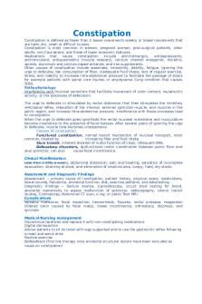
Constipation Constipation is defined as fewer than 3 bowel movements weekly or bowel movements that are hard, dry, small or difficult to pass. Constipation is most common in women, pregnant women, post-surgical patients, older adults, non-Caucasians, and those of lower economic statuses. Medications that cause constipation include anticholinergics, antidepressants, anticonvulsant, antispasmodics (muscle relaxers), calcium channel antagonist, diuretics, opioids, aluminum and calcium-based antacids, and iron supplements. Other causes of constipation include weakness, immobility, debility, fatigue, ignoring the urge to defecate, low consumption of fiber, inadequate fluid intake, lack of regular exercise, stress, and inability to increase intra-abdominal pressure to facilitate the passage of stools for example patients with spinal cord injuries or emphysema (lung condition that causes SOB). Pathophysiology Interference with mucosal secretions that facilitate movement of colon content, myoelectric activity, or the processes of defecation. The urge to defecate in stimulated by rectal distension that then stimulates the inhibitory re4ctoanal reflex, relaxation of the internal, external sphincter muscle, and muscles in the pelvic region, and increase intra-abdominal pressure. Interference with these processes lead to constipation. When the urge to defecate goes ignor3edd the rectal mucosal membrane and musculature become insensitive to the presence of fecal masses. After several years of ignoring the urge to defecate, muscle tone becomes unresponsive. Classes of constipation: Functional constipation, normal transit mechanism of mucosal transport, most common, treated by increasing fiber and fluid intake. Slow transit, inherent disorder of motor function of colon, infrequent BMs. Defecatory disorders, dysfunctional motor coordination between pelvic floor and anal sphincter, can also cause fecal incontinence. Clinical Manifestation Less than 3 BMs a weeks, abdominal distension, pain and bloating, sensation of incomplete evacuation, straining at stool, and elimination of small-volume, lumpy, hard, dry stools. Assessment and Diagnostic Findings Assessment ~ primary cause of constipation, patient history, physical exam, medications, bowel sounds, flatulence, anorectal function, diet, exercise patterns, and labs/testing. Diagnostic Findings ~ Barium enema, sigmoidoscopy, occult stool testing for blood, anorectal manometry to assess malfunction of sphincter, defecography, colonic transit studies, Colonoscopy. Abdominal CT scan, x-ray, or pelvic floor MRI. Complications Valsalva maneuver, fecal impaction, hemorrhoids, fissures, rectal prolapse, megacolon (dilated colon caused by fecal mass), bowel incontinence, orthostasis, dizziness, and syncope. Medical/Nursing management Discontinue laxatives and replace it with non-constipating medications Digital disimpaction Advise patients to sit on toilet with legs supported and to use the gastrocolic reflex following a meal and warm drink Routine exercise Biofeedback (first line therapy once anorectal structural lesions have been excluded as cause on constipation)
Dietary intake of 25-30 grams of fiber a day Patient education Laxatives: bulk forming agents, saline and osmotic agents, lubricants, stimulants, fecal softeners, if enema or rectal suppository is in dire need, glycerin suppositories may be tried.
Diarrhea Diarrhea is an increased frequency of bowel movements (more than 3 per day) with altered consistency (increased liquid) of stool. Diarrhea can be associated with urgency, perineal discomfort, incontinence, nausea, or a combination of these factors. Any condition that increases intestinal secretions, decrease mucosal absorption, or alter motility can produce diarrhea. Causes of diarrhea include viral infections, drugs (ex. Antacids, antibiotics, antihypertensives, antiarrhythmics), adverse effects of chemotherapy, DM, Addison's disease, thyrotoxicosis, lactose intolerance, celiac disease, anal sphincter defect, Zollinger Ellison syndrome, AIDS, and C-Diff (most common HAI in US). Acute diarrhea is self-limiting, lasting 1-2 days; persistent diarrhea typically lasts 2-4 weeks; chronic diarrhea persists for more than 4 weeks and may return periodically. Pathophysiology Acute and persistent diarrheas are either noninflammatory diarrhea (large volume, loose, watery stools) or inflammatory diarrhea (small volume, bloody stool). Organisms implicated include shigella, salmonella, and yersinia species. Types of chronic diarrhea include: Secretory diarrhea, high volume diarrhea caused by increased production of and secretion of water and electrolytes by intestinal mucosa into lumen associated with bacterial toxins or chemotherapeutic agents. Osmotic diarrhea occurs when water is pulled into intestines by osmotic pressure of unabsorbed particles that slow reabsorption of water and it can be caused by lactase deficiency, pancreatic dysfunction, or intestinal hemorrhage. Malabsorptive diarrhea, Inhibited absorption of nutrients. Low albumin levels lead to Intestinal mucosal swelling and liquid stools. Infectious diarrhea results from infectious agents invading the intestinal mucosa. Exudative diarrhea is caused by changes in mucosal integrity, epithelial loss, or tissue destruction by radiation or chemotherapy. Clinical Manifestations Increased frequency and fluid content of stool, abdominal cramps, distension, borborygmus (rumbling or gurgling noise made by the movement of fluid and gas in the intestines), anorexia, thirst, painful spasmodic contractions of the anus, tenesmus (painful straining with a strong urge), dehydration, and electrolyte imbalances. Voluminous, greasy stools indicate intestinal malabsorption. Presence of blood, mucus, and pus in the stool suggest inflammatory enteritis or colitis. Oil droplets on toilet water may suggest pancreatic insufficiency. Nocturnal diarrhea may be a manifestation of diabetic neuropathy. C-Diff infection should be considered in all patients with unexplained diarrhea. Assessment and Diagnostic Findings
Assessment ~ History of present illness, recent travels, diet, stool quality, blood amount if any, frequency, palpate for tenderness, assess mucous membranes, monitor for perineal skin breakdown. Diagnostic Findings ~ CBC, serum levels, urinalysis, endoscopy, barium enema, or routine stool examination for infectious or parasitic organisms, bacterial toxins, blood, fat, electrolytes, and WBC. Complications Dehydration, electrolyte loss (especially of potassium), cardiac dysrhythmias, metabolic acidosis, muscle weakness, paresthesia, hypotension, anorexia, drowsiness, irritant dermatitis, and oliguria. Medical/Nursing Management
Control symptoms Prevent complications Eliminate or treat underlying causes Prevent transmission of infection Monitor diarrheal patterns and characteristics Assess health history, recent travels, and dietary habits Abdominal, skin, perineal area, and mucous membrane assessment Obtain stool samples for testing Increase fluids and foods low in bulk Avoid caffeine, ETOH, dairy, and fatty foods Administer antidiarrheal medications (diphenoxylate with atropine, loperamide) IV fluids for rapid rehydration Monitor electrolyte levels and evidence of dysrhythmias
Peritonitis Inflammation of the serous membrane lining the abdominal cavity and covering the viscera (peritoneum). Peritonitis is a result of bacterial infection, abdominal surgery, trauma, or an inflammation that extends from an organ outside the peritoneal are such as the kidneys or peritoneal dialysis. Peritonitis can be categorized as: Primary peritonitis, also called spontaneous bacterial peritonitis (SBP) that occurs as a spontaneous bacterial infection of ascitic fluid. Most common in liver failure patients. Secondary peritonitis occurs secondary to a perforated abdominal organ with spillage that infects the serous peritoneum. Tertiary peritonitis occurs as a result of superinfection in immunocompromised patients. Pathophysiology Leakage of contents from abdominal organs into the abdominal cavity usually as a result of inflammation, infection, ischemia, trauma, or tumor perforation. Edema of the tissues result, and exudation of fluids develop in a short period of time. Fluid in abdominal cavity increases protein, WBC, cellular debris, and blood amounts in the body. Clinical Manifestations Pain over area, presence of a cause, rebound tenderness, abdominal distension and rigidity, fever (>100F), anorexia, N/V, tachycardia, hypertension then with progression patients may become hypotensive, hypoactive bowel sounds, hypovolemia due to massive amounts of fluid and electrolytes moving from intestinal lumen to the peritoneal cavity and deplete fluid in the vascular space causing shock, and dehydration. Assessment and Diagnostic Findings Assessment ~ Elevated WBC count, decreased hematocrit and hemoglobin levels, and altered potassium, sodium, and chloride electrolyte balances. Diagnostic Findings ~ x-ray may show air and fluid levels and distended bowel loops, Abdominal ultrasound may reveal abscesses (pus filled lumps) and fluid collections, ultrasound-guided aspiration may assist in easier placement of drains, CT scan may show abscess formation, Peritoneal aspiration and C&S studies of aspired fluids may reveal
infection and causative organisms, and MRI may be used to diagnose intra-abdominal abscesses. Medical/Nursing Management Fluid, colloid, and electrolyte replacement (isotonic solution) Analgesics for pain Antiemetics for N/V Intestinal intubation and suction Oxygen therapy via nasal canula, mask, or airway intubation and ventilatory assistance Antibiotics Identify source of infection and control/eliminate it Surgery Drainage of fluids Monitor for s/sx of shock Increase fluids and food intake
Diverticulitis Diverticulum is a saclike herniation of the lining of the bowel that extends through a defect in the muscle layer. Occurs most commonly in the sigmoid colon. Diverticulosis is the presence of multiple diverticula without inflammation or symptoms. Diverticulitis is inflammation and infection of the bowel mucosa caused by bacteria, food, or fecal matter trapped in one or more diverticula (pouch-like herniations in the intestinal wall) Not all clients who have diverticulosis develop diverticulitis. Contributing factors of diverticulitis is aging, constipation, obesity, history of cigarette smoking, regular use of NSAIDs drugs and Tylenol, family history, low fiber and high fat diet, and connective tissue disorders causing weakness in the colon walls. Pathophysiology Bowel content can accumulate in the diverticulum and decompose, causing inflammation and infection. The inflammation of the weakened colonic wall of the diverticulum can cause it to perforate, giving rise to irritability and spasticity of the colon which then leads to diverticulitis. When a patient develops symptoms of diverticulitis, microperforation (series of small holes in the colon) of the colon has occurred. Clinical manifestations Chronic constipation, fistula formation, cramps, narrow stools, intestinal obstruction, diarrhea, nausea, chills or fever, tachycardia, anorexia, bloating, abdominal distention, bleeding, and acute onset of mild to severe pain in the lower left quadrant. If abscess develops, tenderness, palpable mass, fever and leukocytosis occurs.
Assessment and Diagnostic Findings Assessment ~ Colonoscopy visualizes the extent of diverticular disease. CBC reveals an elevated white blood cell count to assist in diagnosis of diverticulitis. Hemoglobin levels should be analyzed for patients with blood in the stool. Urinalysis and urine culture should be analyzed in patients with suspected colovesicular fistulas. Diagnostic Findings ~ Hematocrit and hemoglobin: Decreased, ESR: Increased, WBC: Increased, Stool for occult blood: Can be positive. An abdominal CT scan with contrast agent confirms the diagnosis of diverticulitis and it can also reveal perforation and abscesses. Abdominal x-rays may demonstrate free air under the diaphragm if a perforation has occurred from the diverticulitis. Medical/Nursing Management For severe manifestations (severe pain, high fever), the client is hospitalized, NPO (low fiber diet), and receives nasogastric suctioning, IV fluids, IV antibiotics, and opioid analgesics for pain. Instruct the client who has mild diverticulitis about self-care at home. The client should take medications as prescribed (antibiotics, analgesics, antispasmodics), get adequate rest, drink adequate amounts of fluid, and clear liquids until inflammation subsidies then high-fiber and low-fat diet. Provide the client with instructions to promote normal bowel function and consistency. Individualized exercise program encourages patient to improve abdominal muscle tone. Avoiding foods that trigger diverticulitis attack. Suggests foods that are soft but have increased fiber. Drink adequate fluids.
Surgery: Abscess I&D (incision and drainage to drain the abscess) Resection (removal of inflamed area) Colostomy
Bowel/Intestinal Obstruction Bowel obstruction occurs when blockage prevents the normal flow of intestinal contents through the intestinal tract. Mechanical obstruction: obstruction from pressure on the intestinal walls that occur. (polypoid tumors, intussusception, neoplasm, stenosis, strictures, adhesions, hernias, abscesses, and bezoars). Functional or paralytic obstruction: intestinal musculature cannot prepare the contents along the bowel. This can be a temporary result of manipulation of the bowel during surgery. (amyloidosis, muscular dystrophy, diabetes, Parkinson’s). Bowel obstructions can be defined as partial or complete and simple or strangulated. Partial obstruction is when gas or liquid stool can pass through the point of narrowing, and complete obstruction is when no substance can pass. Contributing factors to intestinal obstruction include crohns disease, radiation therapy, fecal impaction, carcinoma’s, surgical procedures, narcotics, hypokalemia, and diverticulitis.
Small bowel obstruction Clinical manifestations: Crampy colicky pain, passing of blood and mucus but no fecal matter, no flatus, vomiting, reversed peristalsis, signs of dehydration (thirst, drowsiness, malaise, aching, dry mouth), abdominal distention, metabolic alkalosis, hypovolemic shock, and septic shock. Assessment and diagnostic findings: Abdominal x-ray and CT scan findings include abnormal quantities of gas, fluid, or both in the intestines and sometimes collapsed distal bowel. Electrolyte studies in a CBC reveals a picture of dehydration, loss of plasma volume, and possible infection. Medical/Nursing Management: Medical management includes decompression of the bathroom and NG tube, resting the bowel, IV fluids to replace depleted water, sodium, chloride, and potassium. Surgery: Affected portion of bowel is either repaired or removed, laparoscopy or open laparotomy Nursing management is maintaining the function of NG tube, assessing in measuring the NG output, assessing for fluid and electrolyte imbalance is, monitoring nutritional status, reporting discrepancies in I&O, and reporting worsening pain to MD. Complications: Strangulation and tissue necrosis Large bowel obstruction Clinical manifestations: Constipation for weeks, obstruction increasing in size, blood in stool, weakness, weight loss, anorexia, and crampy lower abdominal pain Assessment and diagnostic findings: Abdominal x-ray, CT scan, or MRI findings revealed a distended colon and pinpoint the side of the obstruction. Increased osmolarity and sodium indicates dehydration. Medical/nursing management: Medical management includes the restoration of intravascular volume, correction of electrolyte abnormalities, and NG aspiration and decompression are instituted immediately. NPO.
Surgery: Colonoscopy may be performed to decompress and untwist the bowel. Cecostomy may be performed in patients who are poor surgical risk and urgently need relief from the obstruction. A rectal tube may be use to decompress an area that is lower in the bowel. A colonic stent may be used. Surgical resection to remove the obstructing lesion may be performed and a colostomy may be necessary after this procedure. Nursing Management includes monitoring the patient for symptoms indicating worsening obstruction, provide emotional support and comfort, administering IV fluids and electrolytes as prescribed, prepare patient for surgery, and preoperative education. Assess bowel sound.
Inflammatory Bowel Disease IBD is a group of chronic disorders: Crohn’s disease and ulcerative colitis that result in inflammation or ulceration or both of the bowel. Risk factors include race (Caucasian), living in northern climate/urban area, for crohns, women between ages 20-29 who are current smokers, and for ulcerative colitis, men between ages 15 Dash 30 and those older than 60 that are ex-smokers or non-smokers. Crohn’s Disease A subacute and chronic inflammation of the G.I. tract wall that extends through all layers characterized by periods of remission and exacerbation.
Clinical manifestation: prominent RLQ abdominal pain, scar tissue, abdominal tenderness in spasm, crampy pains after meals, weight loss, malnutrition, secondary anemia, weeping, intraabdominal in anal abscesses, fever, leukocytosis. Chronic symptoms include diarrhea, abdominal pain, excessive fat in the feces, anorexia, weight loss, and nutritional deficiencies, joint disorders, skin lesion, ocular disorders, oral ulcers Assessment and diagnostic findings: Stool studies and barium x-rays showing a string sign indicating constriction in a segment of the terminal ileum. CT scan that highlights bowel wall thickening and mesenteric Edema as well as obstructions, abscesses, and fistula’s is considered more sensitive and diagnosing Crohn’s disease. MRI is equally highly sensitive and specific in terms of identifying pelvic and perineal abscess and fistulas. CBC may show decreased hematocrit and hemoglobin levels, elevated white blood cell count. ESR is usually elevated. Albumin in protein levels may be decreased indicating malnutrition. Treatment: Promote adequate rest, record color, volume, frequency, inconsistency of stools, monitor and prevent fluid deficit, nutrition therapy including high calorie protein, low fiber, no dairy, provide support of care, monitor for complications, and prepare for surgery if above measures are not effective (intestinal transplant). Complications: Intestinal obstruction or stricture formation, perianal disease, fluid and electrolyte in balance, malnutrition, fistula an abscess formation, and colon cancer
Ulcerative Colitis Chronic ulcerative an inflammatory disease of the Mucosa in submucosal layers of the colon and rectum that is characterized by unpredictable. Our mission in exacerbation with bouts of abdominal cramps and bloody or purulent diarrhea. Inflammatory changes typically begin in the rectum and progressed proximately through the colon Clinical manifestations: Diarrhea with passage of mucus, purse, or blood, left lower quadrant abdominal pain, intermittent tenesmus, mild to severe bleeding, pallor, anemia, fatigue, anorexia, weight loss, fever, vomiting, dehydration, cramping, passage of six or more liquid stores each day, hypoalbuminemia, electrolyte imbalance, skin lesions, eye lesions, joint abnormalities, and liver disease. Assessment and diagnostic findings: Abdominal x-ray determines cause of symptoms. Colonoscopy distinguishes ulcerative colitis from other diseases of the colon. Biopsies determine histologic characteristics of the colonic tissue and extent of disease. CT scanning, MRI, and ultrasound studies identify abscesses and perry rectal involvement. Stool study will be positive for blood. Examine for parasites in other microbes. Lab test results revealed decreased hematocrit and hemoglobin levels and elevated white blood cell count, low albumin levels, electrolyte imbalance, elevated see reactive protein levels, and elevated antineutrophil cytoplasmic antibody levels. Treatment:
Promote adequate rest, record color, volume, frequency, and consistency of stools, maintain in P or fat is during acute phase, monitor for dehydr...
Similar Free PDFs

Med Surg GI practice questions
- 23 Pages

Ati gi med surg - Gi study guide
- 53 Pages

Lab6 Med Surg Questions
- 4 Pages

Exam 2 Med Surg 2 Practice Questions
- 16 Pages

med surg review questions
- 7 Pages

ATI - Med-Surg Practice A
- 5 Pages

Med Surg Practice A Lopez
- 6 Pages

Lower GI Practice Questions
- 5 Pages

Final Exam Practice Qs - Med Surg
- 19 Pages
Popular Institutions
- Tinajero National High School - Annex
- Politeknik Caltex Riau
- Yokohama City University
- SGT University
- University of Al-Qadisiyah
- Divine Word College of Vigan
- Techniek College Rotterdam
- Universidade de Santiago
- Universiti Teknologi MARA Cawangan Johor Kampus Pasir Gudang
- Poltekkes Kemenkes Yogyakarta
- Baguio City National High School
- Colegio san marcos
- preparatoria uno
- Centro de Bachillerato Tecnológico Industrial y de Servicios No. 107
- Dalian Maritime University
- Quang Trung Secondary School
- Colegio Tecnológico en Informática
- Corporación Regional de Educación Superior
- Grupo CEDVA
- Dar Al Uloom University
- Centro de Estudios Preuniversitarios de la Universidad Nacional de Ingeniería
- 上智大学
- Aakash International School, Nuna Majara
- San Felipe Neri Catholic School
- Kang Chiao International School - New Taipei City
- Misamis Occidental National High School
- Institución Educativa Escuela Normal Juan Ladrilleros
- Kolehiyo ng Pantukan
- Batanes State College
- Instituto Continental
- Sekolah Menengah Kejuruan Kesehatan Kaltara (Tarakan)
- Colegio de La Inmaculada Concepcion - Cebu


