QA 04 2012 - Practice Questions, Dr. Sesack PDF

| Title | QA 04 2012 - Practice Questions, Dr. Sesack |
|---|---|
| Course | Functional Neuroanatomy |
| Institution | University of Pittsburgh |
| Pages | 3 |
| File Size | 70.4 KB |
| File Type | |
| Total Downloads | 25 |
| Total Views | 129 |
Summary
Practice Questions, Dr. Sesack...
Description
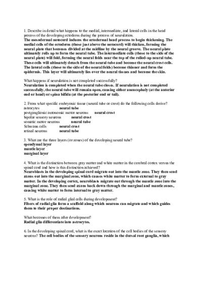
1. Describe in detail what happens to the medial, intermediate, and lateral cells in the head process of the developing ectoderm during the process of neurulation. The mesodermal notocord induces the ectodermal head process to begin thickening. The medial cells of the ectoderm (those just above the notocord) will thicken, forming the neural plate that becomes divided at the midline by the neural groove. The neural plate ultimately rolls up to form the neural tube. The intermediate cells (those to the side of the neural plate) will fold, forming the neural folds near the top of the rolled-up neural tube. These cells will ultimately detach from the neural tube and become the neural crest cells. The lateral cells (those to the side of the neural folds) become thinner and form the epidermis. This layer will ultimately lies over the neural tissues and become the skin. What happens if neurulation is not completed successfully? Neurulation is completed when the neural tube closes. If neurulation is not completed successfully, the neural tube will remain open, causing either anencephaly (at the anterior end or head) or spina bifida (at the posterior end or tail). 2. From what specific embryonic tissue (neural tube or crest) do the following cells derive? astrocytes neural tube postganglionic autonomic motor neurons neural crest bipolar sensory neurons neural crest somatic motor neurons neural tube Schwann cells neural crest retinal neurons neural tube 3. What are the three layers (or zones) of the developing neural tube? ependymal layer mantle layer marginal layer 4. What is the distinction between gray matter and white matter in the cerebral cortex versus the spinal cord and how is this distinction achieved? Neuroblasts in the developing spinal cord migrate out into the mantle zone. They then send axons out into the marginal zone, which causes white matter to form external to gray matter. In the developing cortex, neuroblasts migrate out through the mantle zone into the marginal zone. They then send axons back down through the marginal and mantle zones, causing white matter to form internal to gray matter. 5. What is the role of radial glial cells during development? Fibers of radial glia form a scaffold along which neurons can migrate and which guides them to their proper destinations. What becomes of them after development? Radial glia differentiate into astrocytes. 6. In the developing spinal cord, what is the exact location of the cell bodies of the sensory neurons? The cell bodies of the sensory neurons reside in the dorsal root ganglia, which
develops outside the spinal cord. What is the exact location of the cell bodies of the motor neurons? The cell bodies of the motor neurons are located within the basal plate, which becomes the ventral horn. 7. The sulcus limitans serves as an important divider during the development of the neural tube. What areas are "separated" by the sulcus limitans, and what is the functional significance of this separation? The sulcus limitans divides the dorsal alar plate from the ventral basal plate. In operational terms, this divides structures serving sensory functions from those serving motor functions, respectively. At what point along the neural axis does the sulcus limitans end? The sulcus limitans ends at the top of the mesencephalon and does not extend into the diencephalon. Why does it not extend beyond this point? This is because of the strict definition of the sulcus limitans, namely that it divides the alar plate from the basal plate. The basal plate ends at the mesencephalon because there are no more motor neurons beyond this point. Hence, the sulcus limitans, which no longer separates alar and basal plates, ends. However, there is still a groove in the developing diencephalon and telencephalon that is called the hypothalamic sulcus. This sulcus separates dorsal and ventral portions of the alar plate. 8. Around 4 weeks post-gestation, the embryo exhibits two prominent flexures. What are their names and relative positions? The cephalic flexure lies between the mesencephalon and the rhombencephalon; the cervical flexure lies between the rhombencephalon and the spinal cord. What occurs to each of them during the course of development? The cephalic flexure persists in the adult, whereas the cervical flexure disappears. What is the impact of these changes on the adult nervous system? The disappearance of the cervical flexure allows the spinal cord, medulla and pons to eventually lie along the same axis. The retention of the cephalic flexure causes the nervous system to have a curved longitudinal axis. 9. Name at least one brain structure that derives from the following vesicles: prosencephalon cortex, basal ganglia, thalamus, hypothalamus, pituitary, retina, olfactory bulb metencephalon pontine nucleus, cerebellum rhombencephalon A @ + medulla 10. What is the name of the fluid-filled space within the diencephalon? Within the spinal cord? third ventricle
central canal...
Similar Free PDFs

Sample/practice exam 2012, questions
- 30 Pages

Sample/practice exam 2012, questions
- 11 Pages

DIN 30670 2012-04 EN.pdf
- 37 Pages

Williamson 2012- practice example
- 16 Pages

04-S Functions - Dr. Vaidy
- 11 Pages

Exam 2012, questions
- 4 Pages

Exam 2012, questions
- 8 Pages

Exam 2012, questions
- 1 Pages

Exam 2012, questions
- 16 Pages

Final Exam questions 2012
- 10 Pages

Exam 2012, questions
- 7 Pages
Popular Institutions
- Tinajero National High School - Annex
- Politeknik Caltex Riau
- Yokohama City University
- SGT University
- University of Al-Qadisiyah
- Divine Word College of Vigan
- Techniek College Rotterdam
- Universidade de Santiago
- Universiti Teknologi MARA Cawangan Johor Kampus Pasir Gudang
- Poltekkes Kemenkes Yogyakarta
- Baguio City National High School
- Colegio san marcos
- preparatoria uno
- Centro de Bachillerato Tecnológico Industrial y de Servicios No. 107
- Dalian Maritime University
- Quang Trung Secondary School
- Colegio Tecnológico en Informática
- Corporación Regional de Educación Superior
- Grupo CEDVA
- Dar Al Uloom University
- Centro de Estudios Preuniversitarios de la Universidad Nacional de Ingeniería
- 上智大学
- Aakash International School, Nuna Majara
- San Felipe Neri Catholic School
- Kang Chiao International School - New Taipei City
- Misamis Occidental National High School
- Institución Educativa Escuela Normal Juan Ladrilleros
- Kolehiyo ng Pantukan
- Batanes State College
- Instituto Continental
- Sekolah Menengah Kejuruan Kesehatan Kaltara (Tarakan)
- Colegio de La Inmaculada Concepcion - Cebu


