Skeleton Anatomy Viewer Game questions and answers 2020-2021 worksheet PDF

| Title | Skeleton Anatomy Viewer Game questions and answers 2020-2021 worksheet |
|---|---|
| Author | al top |
| Course | Human Functional Anatomy |
| Institution | Yale University |
| Pages | 3 |
| File Size | 167.1 KB |
| File Type | |
| Total Downloads | 112 |
| Total Views | 151 |
Summary
Answers to skeleton anatomy viewer onlineAnswers to skeleton anatomy viewer onlineAnswers to skeleton anatomy viewer onlineAnswers to skeleton anatomy viewer onlineAnswers to skeleton anatomy viewer onlineAnswers to skeleton anatomy viewer onlineAnswers to skeleton anatomy viewer onlineAnswers to sk...
Description
10/29/21, 11:01 AM
Skeleton Anatomy Viewer Game
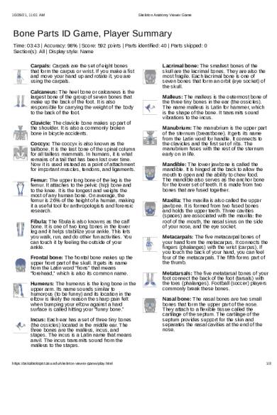
Bone Parts ID Game, Player Summary Time: 03:43 | Accuracy: 98% | Score: 592 points | Parts identified: 40 | Parts skipped: 0 Section(s): All | Display style: Name Carpals: Carpals are the set of eight bones that form the carpus or wrist. If you make a fist and move your hand up and rotate it, you are using the carpals. Calcaneus: The heel bone or calcaneus is the largest bone of the group of seven bones that make up the back of the foot. It is also responsible for carrying the weight of the body to the back of the foot. Clavicle: The clavicle bone makes up part of the shoulder. It is also a commonly broken bone in bicycle accidents. Coccyx: The coccyx is also known as the tailbone. It is the last bone of the spinal column for all tailless mammals. In humans, it is what remains of a tail that has been lost over time. Now it is used instead as a point of attachment for important muscles, tendons, and ligaments. Femur: The upper long bone of the leg is the femur. It attaches to the pelvic (hip) bone and to the knee. It is the longest and weighs the most of any human bone. On average, the femur is 26% of the height of a human, making it a useful tool for anthropologists and forensic research. Fibula: The fibula is also knowns as the calf bone. It is one of two long bones in the lower leg and it helps stabilize your ankle. This lets you walk, run, and do other fun activities. You can touch it by feeling the outside of your ankle. Frontal bone: The frontal bone makes up the upper front part of the skull. It gets its name from the Latin word “frons” that means “forehead,” which is also its common name. Humerus: The humerus is the long bone in the upper arm. Its name sounds similar to humorous (to be funny) and its location in the elbow is likely the reason the sharp pain felt when bumping your elbow against a hard surface is called hitting your “funny bone.” Incus: Each ear has a set of three tiny bones (the ossicles) located in the middle ear. The three bones are the malleus, incus, and stapes. The incus is a Latin name that means anvil. The incus transmits sound from the malleus to the stapes.
https://askabiologist.asu.edu/skeleton-viewer-game/play.html
Lacrimal bone: The smallest bones of the skull are the lacrimal bones. They are also the most fragile. Each lacrimal bone is one of seven bones that form an orbit (eye socket) of the skull. Malleus: The malleus is the outermost bone of the three tiny bones in the ear (the ossicles). The name malleus is Latin for hammer, which is the shape of the bone. It transmits sound vibrations to the incus. Manubrium: The manubrium is the upper part of the sternum (breastbone). It gets its name from the Latin word for handle. It connects to the clavicles and the first set of ribs. The manubrium fuses with the rest of the sternum early on in life. Mandible: The lower jawbone is called the mandible. It is hinged at the back to allow the mouth to open and the ability to chew food. The mandible also serves as the anchor bone for the lower set of teeth. It is made from two bones that are fused together. Maxilla: The maxilla is also called the upper jawbone. It is formed from two fused bones and holds the upper teeth. Three cavities (spaces) are associated with the maxilla: the roof of the mouth, the nasal sinus on the side of your nose, and the eye socket. Metacarpals: The five metacarpal bones of your hand form the metacarpus. It connects the fingers (phalanges) with the wrist (carpus). If you touch the back of your hand, you can feel four of the metacarpals. The fifth forms part of the thumb. Metatarsals: The five metatarsal bones of your foot connect the back of the foot (tarsals) with the toes (phalanges). Football (soccer) players commonly break these bones. Nasal bone: The nasal bones are two small bones that form the upper part of the nose. They attach to a flexible tissue called the cartilage of the septum. The cartilage of the septum provides support for the skin and separates the nasal cavities at the end of the nose.
1/3
10/29/21, 11:01 AM
Occipital bone: Located at the back and lower part of the skull, the occipital bone is a triangleshaped section that connects to the parietal and temporal bones. This bone rests on top of the spinal column. Parietal bone: The two bones that make up the sides and top of the skull are called the parietal bones. Patella: The patella bones are commonly called the kneecaps. The patella moves with the femur and helps protect the front of the knee. It is the largest sesamoid bone (a bone inside of a tendon) in humans. In young athletes, it can sometimes slide out of position and cause extreme pain. Pelvic bone: The two pelvic bones are also called hip bones. Each pelvic bone is made of three bones (the ilium, ischium, and pubis) that fuse together in young adults. The two hip bones, along with the sacrum and coccyx, form the pelvis. Women usually have a wider pelvis than men to allow childbirth. Foot phalanges: The phalanx bones (plural phalanges) form the fingers and toes. Most toes have three phalanx bones, but the big toe only has two. The proximal foot phalanges are closest to the ankle and the distal are closest to the toe tips. The intermediate is in between, but not on the big toe. Phalanges: The phalanx bones (plural phalanges) form the fingers and toes. Most fingers have three phalanx bones, but the thumb only has two. The proximal phalanges are closest to the wrist and the distal are closest to the finger tips. The intermediate is in between, but not on the thumb. Radius: The radius, also called the radial bone, is one of two long bones in the forearm. Its name helps tell what the bone does. It is able to rotate around the other long bone of the forearm. Holding your arm out forward and moving the palm of the hand so that it faces either up or down is an example of how the radius bone rotates. Ribs: This collection of bones has several roles, but maybe their most important is to protect organs such as the heart and lungs. Along with the sternum and the 12 thoracic vertebrae, the ribs make up the rib cage. All the rib bones are connected to the vertebrae at the back, but only some of the ribs are connected to the sternum at the front.
https://askabiologist.asu.edu/skeleton-viewer-game/play.html
Skeleton Anatomy Viewer Game
Sacrum: The sacrum is located at the base of the spine and is made from five fused vertebrae. It connects the last vertebra (L5) and the coccyx (tailbone). The five vertebrae begin as separate bones and start to fuse around the age of 16-18. Generally the sacrum becomes a single bone in the mid-thirties. Scapula: The scapulae (singular scapula) are commonly called the “shoulder blades.” The word “scapula” is actually Latin for “blade.” The muscles and tendons attached to the scapula, such as the four rotator cuff muscles, can be damaged, especially in athletes. Sphenoid bone: This wedge-shaped bone is located just in front of the temporal bone. It gets its name from the Greek word sphenoeides meaning “wedge like.” The sphenoid is one of seven bones that form the orbit (eye socket) of the skull. Stapes: Each ear has a set of three tiny bones (the ossicles). The three bones are the malleus, incus, and stapes. They transfer sound waves in air to fluid-membrane waves. Of these bones, the stapes is the smallest, lightest, and closest to the inner ear. It is shaped like a stirrup. Sternum: The sternum bone is also called the breastbone. Together with the ribs and thoracic vertebrae, the sternum makes up the rib cage. In early development, the sternum has three sections: the manubrium, the body, and the xiphoid process. These sections fuse into one bone. In heart surgery, the sternum may need to be cut lengthwise to gain access to the heart. Tarsals: The tarsal bones are the group of seven bones that make the back part of each foot. The calcaneus (heel bone) is the largest of these bones and is responsible for bearing the weight of the body in the heel of the foot. Teeth: Both bones and teeth are white and hold calcium, phosphorous, and other minerals. But teeth are not bone. Bone is made of collagen, a living and growing tissue. Teeth are even harder than bone and are made of a tissue called dentine that is calcified. Teeth also have an outer layer of enamel that makes them shiny and harder. Temporal bone: There are two temporal bones of the skull, one on either side of the head. Each temporal bone includes an opening for the ear canal. The major nerves of the head and vessels to and from the brain also cross the temporal bone.
2/3
10/29/21, 11:01 AM
Tibia: The tibia is one of the two bones found in the leg that connect the knee with the ankle. It is also called the shin bone. If you touch the front part of your leg below the knee you can feel this bone. The tibia is the second largest bone in the human body and also one of the strongest weight-bearing bones. Ulna: The ulna, or elbow bone, is one of the two bones that are found in the forearm. Bumping the elbow against a hard object can cause a sharp pain that is called bumping your “funny bone” due to how close it is to the humerus bone. The pain from hitting the elbow comes from the ulnar nerve that is just below the skin. Cervical vertebrae: The seven cervical vertebrae are the smallest of the vertebrae. Located just below the head, they are named and numbered C-1 through C-7. Just below the skull is C-1. C-1 (the Atlas) and C-2 (the Axis) connect the skull with the spine. These two bones allow the head to twist. The occipital bone of the skull and C-1 allow the head to nod.
Skeleton Anatomy Viewer Game
Lumbar vertebrae: These 5 vertebrae are the largest of the backbone. They are important for supporting the weight of the body and allowing the body to move. The vertebrae are named and numbered L-1 through L-5. L-1 is the upper-most vertebra. Thoracic vertebrae: The middle section of the backbone is made of the 12 thoracic vertebrae. Along with the ribs and sternum, they make up the rib cage that protects the heart, lungs, and other organs. The vertebrae are commonly named and numbered T-1 through T-12. The closest vertebra to the head is T-1. Xiphoid process: The xiphoid process is one of three parts of the sternum. In younger adults, it is mainly made of cartilage; it usually turns into bone after the age of 15. It is possible to break the xiphoid process, which can cause internal damage. For this reason, care should be taken not to apply pressure to this area when giving CPR. Zygomatic bone: The zygomatic bone is also called the cheekbone. It is one of seven bones that forms the orbit of the skull.
Busy Bones (askabiologist.asu.edu/busy-bones) | Ask A Biologist (askabiologist.asu.edu) | Arizona State University (asu.edu)
https://askabiologist.asu.edu/skeleton-viewer-game/play.html
3/3...
Similar Free PDFs

Axial skeleton quiz answers
- 14 Pages

skeleton answers final
- 45 Pages

6. The Axial Skeleton Worksheet
- 8 Pages

Cell Anatomy Worksheet 1
- 3 Pages
Popular Institutions
- Tinajero National High School - Annex
- Politeknik Caltex Riau
- Yokohama City University
- SGT University
- University of Al-Qadisiyah
- Divine Word College of Vigan
- Techniek College Rotterdam
- Universidade de Santiago
- Universiti Teknologi MARA Cawangan Johor Kampus Pasir Gudang
- Poltekkes Kemenkes Yogyakarta
- Baguio City National High School
- Colegio san marcos
- preparatoria uno
- Centro de Bachillerato Tecnológico Industrial y de Servicios No. 107
- Dalian Maritime University
- Quang Trung Secondary School
- Colegio Tecnológico en Informática
- Corporación Regional de Educación Superior
- Grupo CEDVA
- Dar Al Uloom University
- Centro de Estudios Preuniversitarios de la Universidad Nacional de Ingeniería
- 上智大学
- Aakash International School, Nuna Majara
- San Felipe Neri Catholic School
- Kang Chiao International School - New Taipei City
- Misamis Occidental National High School
- Institución Educativa Escuela Normal Juan Ladrilleros
- Kolehiyo ng Pantukan
- Batanes State College
- Instituto Continental
- Sekolah Menengah Kejuruan Kesehatan Kaltara (Tarakan)
- Colegio de La Inmaculada Concepcion - Cebu


