Skull Foramina PDF

| Title | Skull Foramina |
|---|---|
| Course | Head and Visceral Anatomy |
| Institution | Royal Melbourne Institute of Technology |
| Pages | 6 |
| File Size | 479.8 KB |
| File Type | |
| Total Downloads | 99 |
| Total Views | 139 |
Summary
SKULL FORAMINA WEEKS 1-3...
Description
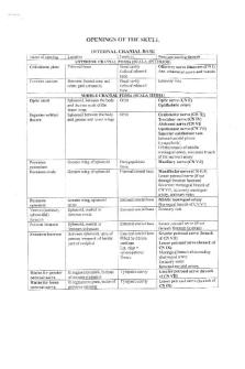
1. Name all those holes in the base of the skull and tell us what goes through each. Cranial fossa
Bone
Foramina
Vessels
Nerves
supraorbital foramen
supraorbital artery, supraorbital nerve supraorbital vein
frontal
anterior cranial fossa
foramen cecum
emissary veins to superior sagittal sinus
-
ethmoid
-
foramina of cribriform plate
-
olfactory nerve bundles (I)
ethmoid
anterior cranial fossa
anterior ethmoidal foramen
ethmoid
anterior cranial fossa
posterior ethmoidal foramen
sphenoid
-
optic canal
frontal
-
anterior ethmoidal artery anterior ethmoidal vein posterior ethmoidal artery posterior ethmoidal vein ophthalmic artery
anterior ethmoidal nerve
posterior ethmoidal nerve optic nerve (II) oculomotor nerve (III) trochlear nerve (IV) lacrimal, frontal and nasociliary branches of ophthalmic nerve (V 1 ) abducent nerve (VI)
sphenoid
middle cranial fossa
superior orbital fissure
superior ophthalmic vein inferior ophthalmic vein
sphenoid
middle cranial fossa
foramen rotundum
-
maxillary nerve (V 2 )
-
incisive foramen/incisive canals
sphenopalatine artery
nasopalatine nerve(V2)
-
greater palatine foramen
-
lesser palatine foramina
-
inferior orbital fissure
maxilla
palatine palatine and maxilla sphenoid and maxilla
greater palatine artery greater palatine vein lesser palatine artery lesser palatine vein inferior ophthalmic veins infraorbital artery
greater palatine nerve
lesser palatine nerve zygomatic nerve and infraorbital nerve of maxillary nerve (V 2 )
infraorbital vein
orbital branches of pterygopalatine ganglion
maxilla
-
infraorbital foramen
infraorbital artery infraorbital vein
infraorbital nerve
sphenoid
middle cranial fossa
foramen ovale
accessory meningeal artery
mandibular nerve (V 3 ) lesser petrosal nerve (occasionally)
sphenoid
middle cranial fossa
foramen spinosum
middle meningeal artery
meningeal branch of the mandibular nerve (V 3 )
sphenoid
temporal
middle cranial fossa middle cranial fossa
internal carotid foramen lacerum artery, artery of pterygoid canal
nerve of pterygoid canal
internal acoustic meatus
labyrinthine artery
facial nerve (VII), vestibulocochlear nerve (VIII)
inferior petrosal sinus, sigmoid sinus
glossopharyngeal nerve (IX), vagus nerve (X), accessory nerve (XI)
temporal
posterior cranial fossa
jugular foramen
occipital
posterior cranial fossa
hypoglossal canal -
hypoglossal nerve (XII)
occipital
posterior cranial fossa
anterior and posterior spinal foramen magnum arteries, vertebral arteries
medulla oblongata
temporal
posterior cranial fossa
stylomastoid foramen
stylomastoid artery facial nerve
2. Review the boundaries of anterior, middle and posterior cranial fossa. Anterior cranial fossa: Anterior limit is the posterior wall of the frontal sinus. The anterior clinoid processes and the planum sphenoidale, which forms the roof of the sphenoid sinus, mark the posterior limit. The frontal bone forms the lateral boundaries. The frontal bone houses the supraorbital foramina, which, along with the frontal sinuses, form 2 important surgical landmarks during approaches involving the anterior skull base. Middle cranial fossa: The greater wing of the sphenoid helps form the anterior limit of the middle skull base. The posterior limit is the clivus. The greater wing of the sphenoid forms the lateral limit as it extends laterally and upward from the sphenoid body to meet the squamous portion of the temporal bone and the anteroinferior portion of the parietal bone. The greater wing of the sphenoid forms the anterior floor of the fossa. The anterior aspect of the petrous temporal bone forms the posterior floor of the middle cranial fossa. The body of the sphenoid makes up the central portion of the middle fossa and houses the sella turcica. Posterior cranial fossa: The posterior skull base consists of primarily the occipital bone, with contributions from the sphenoid and temporal bones. The basal portion of the occipital bone (the basiocciput) and the basisphenoid form the anterior portion of the
posterior skull base. These 2 regions combine to form the midline clivus. The posterior surface of the petrous temporal bone and the lateral aspect of the occipital bone form the lateral wall. The occipital bone also fuses with the mastoid portion of the temporal bone to form the occipitomastoid suture. The petrous portion of the temporal bone and the greater wings of the sphenoid bone are particularly important for identifying structures. The overlying tentorium cerebelli separates the cerebellum from the cerebral hemispheres above, whereas the occipital bone form the lateral walls and floor. 3. List the contents of the superior and inferior orbital fissures. Superior orbital fissure:
superior and inferior divisions of oculomotor nerve (III) trochlear nerve (IV) lacrimal, frontal and nasociliary branches of ophthalmic nerve (V1 ) abducens nerve (VI) superior and inferior divisions of ophthalmic vein. Inferior division also passes through the inferior orbital fissure. sympathetic fibers from cavernous plexus
The nerves passing through the fissure can be remembered with the mnemonic, "Live Frankly To See Absolutely No Insult" - for Lacrimal, Frontal, Trochlear, Superior Division of Oculomotor, Abducens, Nasociliary and Inferior Division of Oculomotor nerve. It is divided into 3 parts from lateral to medial: Lateral Part transmits: lacrimal nerve, frontal nerve, trochlear nerve, meningeal branch of lacrimal artery, anastomotic branch of middle meningeal artery which anastomoses with recurrent branch of the lacrimal artery Middle Part transmits: Upper and lower divisions of the oculomotor nerve, nasociliary nerve between the two divisions of oculomotor nerve and abducent nerve Medial Part transmits: Superior ophthalmic vein and sympathetic nerves from the plexus around internal carotid artery Inferior orbital fissure: infraorbital nerve (a branch of V2), infraorbital artery (a branch of the third part of the maxillary artery) Anatomy of the left orbital apex, highlighting the extraocular muscle origins and the contents of the superior orbital fissure. Key: LPS, levator muscle; SR, superior rectus; LR, lateral rectus; IR, inferior rectus; MR, medial rectus; SO, superior oblique; SOV, superior ophthalmic vein; III sup , superior division of oculomotor nerve; III inf , inferior division of oculomotor nerve; IOF, inferior orbital fissure....
Similar Free PDFs

Skull Foramina
- 6 Pages

Hominid Skull
- 3 Pages

Openings of the skull
- 5 Pages

Anterior Skull Base Tumors
- 17 Pages

Skull and trunk notes- sheetal
- 74 Pages
Popular Institutions
- Tinajero National High School - Annex
- Politeknik Caltex Riau
- Yokohama City University
- SGT University
- University of Al-Qadisiyah
- Divine Word College of Vigan
- Techniek College Rotterdam
- Universidade de Santiago
- Universiti Teknologi MARA Cawangan Johor Kampus Pasir Gudang
- Poltekkes Kemenkes Yogyakarta
- Baguio City National High School
- Colegio san marcos
- preparatoria uno
- Centro de Bachillerato Tecnológico Industrial y de Servicios No. 107
- Dalian Maritime University
- Quang Trung Secondary School
- Colegio Tecnológico en Informática
- Corporación Regional de Educación Superior
- Grupo CEDVA
- Dar Al Uloom University
- Centro de Estudios Preuniversitarios de la Universidad Nacional de Ingeniería
- 上智大学
- Aakash International School, Nuna Majara
- San Felipe Neri Catholic School
- Kang Chiao International School - New Taipei City
- Misamis Occidental National High School
- Institución Educativa Escuela Normal Juan Ladrilleros
- Kolehiyo ng Pantukan
- Batanes State College
- Instituto Continental
- Sekolah Menengah Kejuruan Kesehatan Kaltara (Tarakan)
- Colegio de La Inmaculada Concepcion - Cebu


