Stroke pathology : Ischemia & hemorrhagic types PDF

| Title | Stroke pathology : Ischemia & hemorrhagic types |
|---|---|
| Author | Anonymous User |
| Course | Medicine and surgery |
| Institution | University of Otago |
| Pages | 18 |
| File Size | 1.6 MB |
| File Type | |
| Total Downloads | 4 |
| Total Views | 31 |
Summary
Nervous system stroke pathologysIschemic stroke 3i. Global cerebral ischemia 3ii. Focal cerebral ischemia / ischemic infarcts 4iii. Haemorrhagic infarct or non haemorrhage in fact : 5Localising lesions : 6Changes in brain tissue 7gross appearance : Ischemic infarcts : 7Necrotic Inflammation &...
Description
Nervous system stroke pathology Ischemic stroke
3
i. Global cerebral ischemia
3
ii. Focal cerebral ischemia / ischemic infarcts
4
iii. Haemorrhagic infarct or non haemorrhage in fact :
5
Localising lesions :
6
Changes in brain tissue
7
gross appearance : Ischemic infarcts :
7
Necrotic Inflammation & vasogenic oedema
8
Microscopic appearance :
9
Gliosis
11
Hemorrhagic stroke
12
Hypertensive cerebrovascular disease
12
i. Hypertensive intraparenchymal haemorrhages
14
ii. Subarachnoid haemorrhage
15
s"
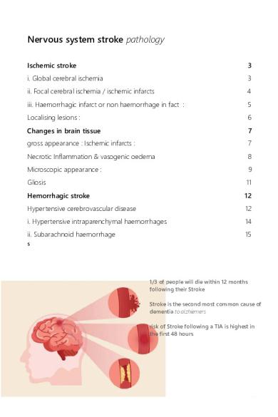
1/3 of people will die within 12 months following their Stroke Stroke is the second most common cause of dementia to alzhiemers risk of Stroke following a TIA is highest in the first 48 hours
Stroke can be defined as any vascular lesion that reduces cerebral blood flow (CBF) to a specific region of the brain, retina, or spinal cord, causing acute neurologic impairment that lasts > 24 hours i.
ischemic stroke : Hypoxic ischemic infarction of the brain caused by occlusion or stenosis
ii.
hemorrhagic stroke caused by vascular rupture causing Intracerebral / intraparenchymal and subarachnoid haemorrhages
This is the third largest killer in NZ age dependent
Common types of ischemic stroke : 1.
Global cerebral ischemia : hypoxic ischemic encephalopathy
2. Focal cerebral ischemia : ischemic infarction caused by thrombosis or embolism
Common causes of hemorrhagic stroke are : 1. Hypertensive intraparenchymal haemorrhage 2. Ruptured aneurysm : subarachnoid haemorrhage
C
Ischemic stroke Neuronal injury form lack of oxygenation and nutritional support -> damage -> death But also accumulation of waste leading to cell damage and death
i. Global cerebral ischemia Generalised decrease in cerebral perfusion due to Shock or hypoxia • Clinical outcome depends on the severity of the insult, ranging from reversible mild injury to severe generalised neuronal death leading to brain death • ie. Cardiovascular shock from cardiac arrest , sepsis and hypotension
Vulnerable areas : • Some areas of the brain are more vulnerable to ischemic injuries than others, including the • hippocampus : • purine cells of the cerebellum : • Watershed areas regions between arterial perfusion territories ie. the most distal reaches of arterial blood supply • ACA water shed area : medially, between the MCA and ACA territory • MCA watershed areas : inferior margin of the temporal lobe
ii. Focal cerebral ischemia / ischemic infarcts Obstruction of blood supply to a localised area of the brain for a long enough period of time can cause ischemic infarction •
Embolism to the brain usually form a thrombus or thromboembolism
•
Thrombosis of vertebral vessels due to atherosclerosis
• Large vessels ie. middle cerebral artery • Small penetrating vessels ie. those supplying the basal ganglia, thalamus and internal capsule note these deeper vessels are more usually associated with hypertension disease
Thrombotic emboli sources : • The heart • Mural thrombus complicating myocardial infarction • Thrombosis complicating atrial fibrillation • Thrombosis complicating vascular diseases ie. rheumatic fever • Aorta & carotid arteries • Thrombosis developing over atheromatous plaques : tiny emboli lead to transient ischemic attacks or ministrokes • Paradoxical emboli • Occur in which thromboembolic from the venous circulation may pass though a cardiac septal effect such as patent foramen scale into the arterial circulation and emboli to the brain • Other forms of embolism are : air embolism , fat embolism, and amniotic fluid embolism
iii. Haemorrhagic infarct or non haemorrhage in fact : Haemorrhages (usually small petechial) secondary to the infarction Hemorrhagic RED infarct : • Typically associated with embolic events • Characterised by multiple sometimes confluent petechial haemorrhages • Later with necrotic tissue • Haemorrhage is due to reperfusion of damaged tissue through : 1. Collateral circulation 2. Following fibrinolysis of thromboembolic material in the occluded vessels and leakage of blood through necrotic vessels
Notice red : micro hemorrhages into the necrotic ischaemic tissue due to reperfusion though damaged necrotic vessels Acute below: notice the Red blood cells
Non hemorrhagic pale / band infarcts : • Usually associated with thrombosis
Localising lesions :
1.
The internal carotid arteries : 2 1.
Anterior cerebral arteries : branches out anteriorly from ICA 1.
Supplies the medial 2/3 surface of the cerebral hemispheres
2. Middle cerebral artery branches laterally from the ICA 1.
All of the lateral surface of the cerebral hemispheres
2. The internal capsule
2. The vertebral arteries : 2 1.
The basilar artery : 1
2. The posterior cerebral arteries : PCA 1.
Posterior 1/3 of the medial surface of the cerebral hemispheres
2. Occipital lobes 3.
inferior temporal lobe
4. superiors brainstem
1.
The hippocampus Z
For a lateral and anterior medial lesion consider the internal carotid artery
Changes in brain tissue gross appearance : Ischemic infarcts : Liquefactive necrosis : the type of hypoxic ischemic necrosis that occurs in the CNS unlike coagulative necrosis where tissue outline is preserved, this type if characterised by digestion of dead tissue into a liquid mass, there is no healing by fibrosis there is no Fibroblasts in the brain (in the exception of an abscess with fibroblast & immune cell infiltration)
Acute • 0-6 hours : • almost undetectable macroscopically : except if its hemorrhagic • 48 hours • : blurring of grey & white matter interface : • the infarct is pale, soft, swollen and ill defined : extensive vasogenic oedema due to disruption of BBB and passage of fluid into extracellular space Later on : • 2-10 days •
the area of the brain infarct becomes gelatinous and friable:
• Well defined border • From 10 days to 3 weeks… forever ! • Tissue breakdown • Liquefaction leaving cystic spaces (fluid filled cavities) • Lined by gliosis
Necrotic Inflammation & vasogenic oedema
The necrotic process will trigger an inflammatory effect that disrupts the BBB to make it more permeable
• Inflammation causes an increase in vascular permeability • Combined with a loss of structural integrity that also causes an increase in vascular permeability of blood vessels due to infarction leads to the extravasation / leaving of fluid known as vasogenic oedema • Over time this may raise ICP and cause brain herniation • Duret haemorrhages : They are due to tearing of penetrating veins and arteries supplying the upper brainstem during progression of transtenotiral uncle herniation seen consequent to raised ICP due to vasogenic oedema
CT appearance :
A. Day of infarct : CT no appearance B. Day of infarct, water weighted in T2 MRI shows areas of edema in region of middle cerebral artery C. Days later, edema shows up as hypotenuse in CT images
Microscopic appearance :
• Early (...
Similar Free PDFs

Class 11 Hemorrhagic Stroke
- 3 Pages

Ischemia Intestinale
- 12 Pages

Pathology
- 26 Pages

stroke
- 11 Pages

SAP STROKE
- 22 Pages

Anaphylaxis - Pathology
- 9 Pages

Oral pathology
- 15 Pages

Cardiovascular Pathology Table
- 21 Pages

Fundamentals of Pathology Pathoma
- 215 Pages

MCQs in Oral Pathology
- 171 Pages

MAKALAH STROKE
- 25 Pages

surveilen stroke
- 14 Pages

PATHOLOGY - EDEMA
- 15 Pages

PNPK Stroke
- 151 Pages
Popular Institutions
- Tinajero National High School - Annex
- Politeknik Caltex Riau
- Yokohama City University
- SGT University
- University of Al-Qadisiyah
- Divine Word College of Vigan
- Techniek College Rotterdam
- Universidade de Santiago
- Universiti Teknologi MARA Cawangan Johor Kampus Pasir Gudang
- Poltekkes Kemenkes Yogyakarta
- Baguio City National High School
- Colegio san marcos
- preparatoria uno
- Centro de Bachillerato Tecnológico Industrial y de Servicios No. 107
- Dalian Maritime University
- Quang Trung Secondary School
- Colegio Tecnológico en Informática
- Corporación Regional de Educación Superior
- Grupo CEDVA
- Dar Al Uloom University
- Centro de Estudios Preuniversitarios de la Universidad Nacional de Ingeniería
- 上智大学
- Aakash International School, Nuna Majara
- San Felipe Neri Catholic School
- Kang Chiao International School - New Taipei City
- Misamis Occidental National High School
- Institución Educativa Escuela Normal Juan Ladrilleros
- Kolehiyo ng Pantukan
- Batanes State College
- Instituto Continental
- Sekolah Menengah Kejuruan Kesehatan Kaltara (Tarakan)
- Colegio de La Inmaculada Concepcion - Cebu

