The Oculomotor Nerves (III) PDF

| Title | The Oculomotor Nerves (III) |
|---|---|
| Course | Human Anatomy and Physiology with Lab I |
| Institution | The University of Texas at Dallas |
| Pages | 1 |
| File Size | 39 KB |
| File Type | |
| Total Downloads | 93 |
| Total Views | 136 |
Summary
The Oculomotor Nerves (III)...
Description
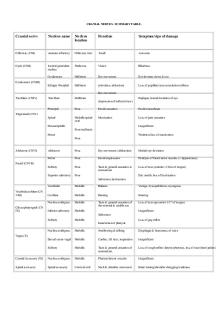
The Oculomotor Nerves (III) ■ Primary function: Motor (eye movements) ■ Origin: Midbrain ■ Pass through: Superior orbital fissures of sphenoid pp. 213, 220, 222, 225 ■ Destination: Somatic motor: superior, inferior, and medial rectus muscles; inferior oblique; levator palpebrae superioris. Visceral motor: intrinsic eye muscles The midbrain contains the motor nuclei controlling the third and fourth cranial nerves. Each oculomotor nerve (III) innervates four of the six extrinsic muscles that move the eye, and the levator palpebrae superioris, which raises the upper eyelid (see Figure 14–21). On each side of the brain, CN III emerges from the ventral surface of the midbrain and penetrates the posterior wall of the orbit at the superior orbital fissure. Individuals with damage to this nerve often complain of pain over the eye, droopy eyelids, and double vision, because the movements of the left and right eyes cannot be coordinated properly. The oculomotor nerve also delivers preganglionic autonomic fibers to neurons of the ciliary ganglion. These neurons control intrinsic eye muscles. These muscles change the diameter of the pupil, adjusting the amount of light entering the eye. They also change the shape of the lens to focus images on the retina. The Trochlear Nerves (IV) ■ Primary function: Motor (eye movements) ■ Origin: Midbrain ■ Pass through: Superior orbital fissures of sphenoid pp. 213, 219, 222, 225 ■ Destination: Superior oblique A trochlear (TRO . K-le . -ar; trochlea, a pulley) nerve (IV), the smallest cranial nerve, innervates the superior oblique of each eye (Figure 14–21). The trochlea is a pulley-shaped, ligamentous sling. Each superior oblique passes through a trochlea on its way to its insertion on the surface of the eye. An individual with damage to cranial nerve IV or to its nucleus has difficulty looking down and to the side. The Abducens Nerves (VI) ■ Primary function: Motor (eye movements) ■ Origin: Pons ■ Pass through: Superior orbital fissures of sphenoid pp. 213, 219, 222, 225 ■ Destination: Lateral rectus The abducens (ab-DU . -senz) nerves (VI) innervate the lateral rectus, the sixth pair of extrinsic eye muscles. Contraction of the lateral rectus makes the eye look to the side. In essence, the abducens causes abduction of the eye. Each abducens nerve emerges from the inferior surface of the brainstem at the border between the pons and the medulla oblongata (Figure 14–22). Along with the oculomotor and trochlear nerves from that side, it reaches the orbit through the superior orbital fissure....
Similar Free PDFs

The Oculomotor Nerves (III)
- 1 Pages

Limb Nerves
- 4 Pages

Cranial Nerves
- 2 Pages

Origin of the Cranial Nerves
- 5 Pages

Plexus Nerves
- 2 Pages

Cranial Nerves
- 2 Pages

Cranial Nerves Worksheet
- 2 Pages

Cranial Nerves Summary
- 4 Pages

20. Cranial Nerves
- 1 Pages

Ch17 Spinal Cord Nerves
- 4 Pages

Spinal Nerves SI - Hargroder
- 4 Pages

Case MS Wearing Nerves AP1
- 4 Pages
Popular Institutions
- Tinajero National High School - Annex
- Politeknik Caltex Riau
- Yokohama City University
- SGT University
- University of Al-Qadisiyah
- Divine Word College of Vigan
- Techniek College Rotterdam
- Universidade de Santiago
- Universiti Teknologi MARA Cawangan Johor Kampus Pasir Gudang
- Poltekkes Kemenkes Yogyakarta
- Baguio City National High School
- Colegio san marcos
- preparatoria uno
- Centro de Bachillerato Tecnológico Industrial y de Servicios No. 107
- Dalian Maritime University
- Quang Trung Secondary School
- Colegio Tecnológico en Informática
- Corporación Regional de Educación Superior
- Grupo CEDVA
- Dar Al Uloom University
- Centro de Estudios Preuniversitarios de la Universidad Nacional de Ingeniería
- 上智大学
- Aakash International School, Nuna Majara
- San Felipe Neri Catholic School
- Kang Chiao International School - New Taipei City
- Misamis Occidental National High School
- Institución Educativa Escuela Normal Juan Ladrilleros
- Kolehiyo ng Pantukan
- Batanes State College
- Instituto Continental
- Sekolah Menengah Kejuruan Kesehatan Kaltara (Tarakan)
- Colegio de La Inmaculada Concepcion - Cebu


