Amniotic Fluid Paper - N/A PDF

| Title | Amniotic Fluid Paper - N/A |
|---|---|
| Course | Medical Technology |
| Institution | Far Eastern University |
| Pages | 3 |
| File Size | 114.5 KB |
| File Type | |
| Total Downloads | 199 |
| Total Views | 656 |
Summary
Download Amniotic Fluid Paper - N/A PDF
Description
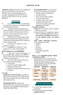
- AMNIOTIC FLUID AMNIOTIC FLUID is formed from the metabolism of fetal urine and fetal cell.It is also subjected to laboratory testing. And these laboratory tests are essential in monitoring FETAL MATURITY and SURVIVAL RATE OF FETUS in cases of FETAL DISTRESS. PHYSIOLOGY
It is found in the AMNION (a membranous sac composed of a single layer of cuboidal epithelial cells that surrounds the fetus) The primary functions of the fluid are to provide a protective cushion for the fetus, allow fetal movement, stabilize the temperature to protect the fetus from extreme temperature changes, and to permit proper lung development. A balance between the production of fetal urine and lung fluid and the absorption from fetal swallowing and intramembranous flow regulates the volume of amniotic fluid. Usual range of its volume at 12 weeks gestation is 25-50 mL; Usual range of its volume at 37 weeks gestation where it peaks at 800-1200 mL.
HOW IS AMNIOTIC FLUID FORMED? It starts at the 1st trimester of pregnancy = 35 mL of amniotic fluid is produce by the AMNION and PLACENTA that is derived from the maternal circulation (maternal plasma). Water and solutes exchange between the fetus and the amniotic fluid through o Fetal urination o Intestinal absorption following fetal swallowing of amniotic fluid o Capillary exchange in the pulmonary system as ALVEOLI. VOLUME AMNIOTIC FLUID VOLUME is regulated by a balance between production of fetal urine and lung fluid and the absorption from fetal swallowing and intramembranous flow. POLYHYDRAMNIOS associated with DECREASED fetal swallowing and often indicates fetal distress and congenital fetal malformation in Neutral Tube Defect (NTD).
OLIGOHYDRAMNIOS is associated with increased fetal swallowing with a possible; o Indication urinary tract deformities o Membrane leakage o Donor twin transfusion syndrome o Congenital malformation o Premature rupture of the amniotic membrane and umbilical cord compression (resulting in decelerated heart rate and fetal death) Chemical Composition Placenta is the ultimate source of amniotic fluid water and solutes. Amniotic fluid has a composition similar to that of the maternal plasma and contains a small amount of sloughed fetal cells from the skin, digestive system, and urinary tract Fluid contains biochemical substances that are produced by the fetus o Bilirubin o Lipids o Enzymes o Electrolytes o Nitrogenous compounds o Proteins HOW IS IT DIFFERENTIATED FROM MATERNAL URINE? Differentiation between amniotic fluid and maternal urine may be necessary to determine possible premature membrane rupture or accidental puncture of the maternal bladder during specimen collection. CRITERI MATERNAL AMNIOTI A URINE C FLUID Creatinine > 10 mg/dL < 3.5 mg/dL Urea > 300 mg/dL < 30 mg/dL Glucose Essentially More NEGATIVE associated Protein Essentially 2-8 g/L NEGATIVE Fern’s test NEGATIVE POSITIVE SPECIMEN COLLECTION, PROCESSING AND ANALYSIS Amniotic fluid is reflective of fetal development. The collection is performed through AMNIOCENTESIS.
It is obtained by NEEDLE ASPIRATICYellow or amber colored fluid: Hemolytic disease of the newborn (HDN). into the amniotic sac. Dark green fluid: AMNIOCENTESIS can either be o Presence of meconium TRANSABDOMINAL (much preferred) or Dark-red-brown fluid: VAGINAL. o Fetal death Typically, Amniocentesis is performed after 14 weeks gestation. However, one must consider the B. TRANSPARENCY purpose of doing the procedure and when it is done. Amniotic fluid would usually have a SLIGHT 15-18 weeks gestation = detection of neural TO MODERATE TURBIDITY. tube defect. TAKE NOTE: FIRST TRIMESTER week 1 to week 12 of pregnancy SECOND TRIMESTER week 13 to week 27 of pregnancy LAST TRIMESTER week 28 to week 40 of pregnancy SPECIMEN HANDLING A. For Fetal Lung Maturity Test (FLM) Specimen must be placed one ice for delivery to the laboratory and refrigerated BEFORE testing within 72 HOURS. The specimen may or may not be centrifuged. B. For Cytogenetic studies The specimen should be maintained at room temperature or body temp. to prolong the life of cells needed for analysis. For CELL CULTURE = 140 x g centrifugation is required. C. For Chemical assay There is a need to separate cellular elements from debris as soon as possible to prevent distortion of chemical constituents DETERMINATION OF PHOSPHOLIPI = centrifugation of 500 to 100 x g for 5 minutes is required.
COLOR AND TRANSPARENCY Normal amniotic fluid is CLEAR and may exhibit slight to moderate turbidity from cellular debris, particularly in later stages of fetal development. A. COLOR Blood-streaked fluid may be present as the result of a: o Traumatic tap o Abdominal trauma o Intra-amniotic hemorrhage
CHEMICAL EXAMINATION A. Test for Fetal age 1. Amniotic fluid creatinine determination It is done by utilizing serum methodJaffe’s reaction. Normal level = ranges between 1.5 and 2.0 mg/dL. B. Test for Fetal Distress 1. Pigment analysis – color variation association i. Hemoglobin ii. Bilirubin analysis In normal pregnancy = bilirubin in amniotic fluid is LOW and UNDETECTABLE at 10-30 mg/dL. SPECTROPOTOMETRIC ANALYSIS = measuring of amniotic fluid bilirubin. 2. Alpha-fetoprotein (AFP) test Measurement of amniotic fluid AFP levels is indicated when maternal serum levels are elevated. 3. Acetylcholinesterase level It is an indication of neural tube defect A confirmatory test after an elevated amniotic fluid AFP level 4. Bacterial count Concentration of bacteria present should be >50/µL of amniotic fluid. C. Test for Fetal Lung Maturity (FLM) 1. Lecithin-Sphingomyelin ratio LECITHIN = The primary component of surfactants (phospholipids, neutral, lipids, and protein). It is produced at a relatively low and constant rate until the 35th week of gestation. SPHINGOMYELIN = a lipid produced at a constant rate after 26 weeks of gestation L/S ratio prior to 35 weeks is usually 2 = indicates MATURITY...
Similar Free PDFs

Amniotic Fluid Paper - N/A
- 3 Pages

Othello playlist - NA/NA
- 2 Pages

Fluid Mechanics
- 796 Pages

fluid & electrolytes
- 7 Pages

Fluid Mechanics
- 8 Pages

Fluid Mechanics
- 260 Pages

fluid mechanics
- 8 Pages

Serous- Fluid
- 7 Pages

Fluid Mechanics, Lecture 4
- 27 Pages

Fluid Mechanics - External Flow
- 7 Pages

Fluid Mechanics Cheat Sheet
- 3 Pages

Fluid Electrolytes worksheet
- 4 Pages

Basic fluid properties
- 3 Pages
Popular Institutions
- Tinajero National High School - Annex
- Politeknik Caltex Riau
- Yokohama City University
- SGT University
- University of Al-Qadisiyah
- Divine Word College of Vigan
- Techniek College Rotterdam
- Universidade de Santiago
- Universiti Teknologi MARA Cawangan Johor Kampus Pasir Gudang
- Poltekkes Kemenkes Yogyakarta
- Baguio City National High School
- Colegio san marcos
- preparatoria uno
- Centro de Bachillerato Tecnológico Industrial y de Servicios No. 107
- Dalian Maritime University
- Quang Trung Secondary School
- Colegio Tecnológico en Informática
- Corporación Regional de Educación Superior
- Grupo CEDVA
- Dar Al Uloom University
- Centro de Estudios Preuniversitarios de la Universidad Nacional de Ingeniería
- 上智大学
- Aakash International School, Nuna Majara
- San Felipe Neri Catholic School
- Kang Chiao International School - New Taipei City
- Misamis Occidental National High School
- Institución Educativa Escuela Normal Juan Ladrilleros
- Kolehiyo ng Pantukan
- Batanes State College
- Instituto Continental
- Sekolah Menengah Kejuruan Kesehatan Kaltara (Tarakan)
- Colegio de La Inmaculada Concepcion - Cebu


