BIOL-1106 - Topic II - Review and Concept Maps PDF

| Title | BIOL-1106 - Topic II - Review and Concept Maps |
|---|---|
| Author | M.R. Smith |
| Course | Principles of Biology |
| Institution | Virginia Polytechnic Institute and State University |
| Pages | 8 |
| File Size | 501.4 KB |
| File Type | |
| Total Downloads | 57 |
| Total Views | 148 |
Summary
Concept maps of information to know for the second test for BIOL-1106....
Description
Daniel T. Eisert BIOL-1106: Principles of Biology
Topic II: Animal Anatomy TEST: July 25, 2017
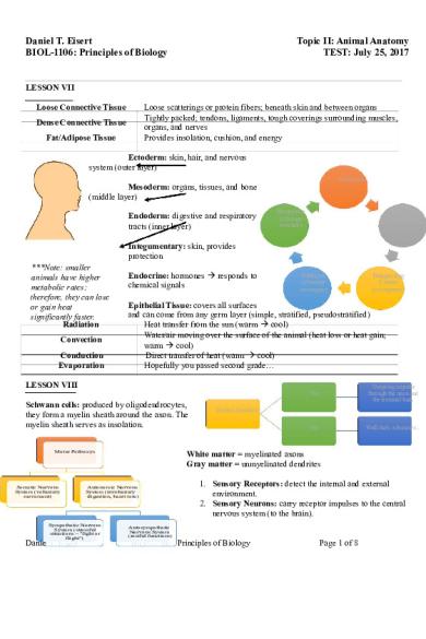
LESSON VII Loose scatterings or protein fibers; beneath skin and between organs Tightly packed; tendons, ligaments, tough coverings surrounding muscles, organs, and nerves Provides insolation, cushion, and energy
Loose Connective Tissue Dense Connective Tissue Fat/Adipose Tissue
Ectoderm: skin, hair, and nervous system (outer layer) Stimulus
Mesoderm: organs, tissues, and bone (middle layer) Endoderm: digestive and respiratory tracts (inner layer)
Response (change worker)
Sensor (monitor)
Integumentary: skin, provides protection ***Note: smaller animals have higher metabolic rates; therefore, they can lose or gain heat significantly faster. Radiation Convection Conduction Evaporation
Endocrine: hormones responds to chemical signals
Effector (change manager)
Epithelial Tissue: covers all surfaces and can come from any germ layer (simple, stratified, pseudostratified) Heat transfer from the sun (warm cool) Water/air moving over the surface of the animal (heat loss or heat gain; warm cool) Direct transfer of heat (warm cool) Hopefully you passed second grade…
LESSON VIII Schwann cells: produced by oligodendrocytes, they form a myelin sheath around the axon. The myelin sheath serves as insolation. Motor Pathways
Somatic Nervous System (voluntary movement)
Danie
Yes
Outgoing impulse through the axon and the terminal buds
No
Well that's a bummer...
Excited Impulses?
White matter = myelinated axons Gray matter = unmyelinated dendrites
Autonomic Nervous System (involuntary digestion, heart rate)
Sympathetic Nervous System (stressful situations ~ "fight or flight")
Integrating Center (compares)
1. Sensory Receptors: detect the internal and external environment. 2. Sensory Neurons: carry receptor impulses to the central nervous system (to the brain).
Autosympathetic Nervous System (restful functions)
Principles of Biology
Page 1 of 8
3. Interneurons: make signals between neurons and integrate those signals; subsequently, they are passed onto the motor neurons. 4. Motor Neurons: carry impulses to effectors (typically a muscle or a gland; from the brain). Somatic NS Responds to convious control, reflexes Motor neuron cell bodies in the CNS Axons extend from the CNS to the effector Axons are highly myleinated Skeletal muscle effectors Increased heart rate, dialated pupils, increased blood flow
Autonomic NS Unconcious/involuntary Two neurons to reach the effector (pre and post) Little or no myleination Stimulatory/inhibitory effect Cardiac and smooth muscle glands are effectors Increase gastric activity, constrict bronchioles
Left Brain Controls right side Lang., math, intrapersonal space Parasynthetic Approach 1. area, Broca’s Wernicke’s area, Exher’s hand area
Right Brain Controls left side Survival, emotional, extrapersonal space Synthetic drive Avoidance
Frontal lobe: executive function, motor cortex, higher order thinking. 2. Parietal lobe: body sensations, math, spatial analysis. 3. Occipital lobe: visual cortex 4. Temporal lobe: auditory complex
Sympathetic Division Stressful situations (lots of energy needed) Nerves originate in the middle portion of the spinal cord Long postganglionic neurons Sympathetic ganglia located outside of the spinal cord
Parasynthetic Division Relaxing (low body energy) Most nerves originate in the brain stem or some regions of the spina cord Long preganglionic neurons Ganglia located near the effector organ
Welcome to the Spinal Cord! ~ Bypassing the Brain (Somatic NS Reflex) Meninges: connective tissue membranes which cover the spinal cord and brain. Gray Matter = inner zone; white matter = myelinated axons of sensory and motor neurons. ***Note: most reflexes are not monosynaptic, for impulses must pass more than one synapse within the gray matter. It is advantageous for reflexes to be myelinated to increase speed and efficiency. Nerves: bundles of axons Passes CNS carries Ganglia: clusters of cell bodies outside of the PNS: Receives information responses to CNS. information
to CNS
the effectors
Preganglionic: in the spinal cord Postganglionic: cell body in a nerve ganglion outside of the CNS transmits signal to an effector (result is either stimulator OR inhibitory).
Inside the cell... More Negative (because of membrane transport proteins) More K Less Na
Autonomic NS Reflex (two motor neurons)
LESSON IX 1. Impulse: the ability to create electric charge differences across the plasma membrane of the neuron.
Hindbrain
Midbrain
Forebrain
Connects the brain to t c r
Receives and
Relay center for
Outside the cell... Less Negative Less K More aniel T.Na Eisert
BIOL-1106: Principles of Biolog
2. As sodium leaves the cell, it allows for the building of potassium ions which dephosphorylates the protein, and subsequently, two potassium ions enter. ***Note: the voltage gated K+ gate is always closed during the resting period; the leaky K+ channels are still open, for they are not voltage gated. ***Note: small changes in membrane potential are referred to as a graded potential. Axon Hillock
1. Electrical synapses: utilize gap junctions to directly transmit action potentials from one cell to another. 2. Chemical synapses: predominant in vertebrates; neurotransmitters are released as ligands from the presynaptic cell and then bound to the postsynaptic cell; they carry messages across the synapse. 3. Presynaptic cell: transmits action potentials to the synapse. 4. Postsynaptic cell: receives the signal from the presynaptic cell
Propagation: the movement of an action potential down the axon Influx of Na+ ions during the rising phase depolarize adjacent sections of membrane to the threshold.
Saltatory Conduction: jumping of action potentials between nodes of Ranvier.
Case Study ~ Drug Addiction: reuptake inhibition by cocaine decrease in the number of receptor proteins. More cocaine is required to feel “good” or eventually just feel normal.
Presynaptic Cell: transmits action potentials. Graded potential is created. If high enough, another A.P. is created.
Action Cell
Channels are opened (Na + or others).
Chemical Synapses
Postsynaptic cell
Danielreceives T. Eisert the proteins.
Action potential reaches the end of the presynaptic neuron.
Calcium ions
BIOL-1106: Principles of Biology
Page 3enter ofgated 8via voltage ion channels.
Influx causes fusion of synaptic vessicles with the plasma membrane.
Relation of neurotransmitter s into the synaptic cleft.
LESSON X Statocysts Motor Receptors Utricle Motor Receptors Saccule Motor Receptors
Mechanoreceptors
Touch, pain, pressure, hearing
Allows invertebrates to detect gravity and motion. Allows vertebrates to detect horizontal motion. Allows vertebrates to detect vertical motion. 1. Inner Ear: receptors for sound waves. Nociceptor: pain, tissue damage, free nerve 2. Middle & Outer Ear: converts sound waves endings (concentrated where injury is likely to occur) into motion. ***Note: in the ear, more stereocilia lets the K+ ions Thermoreceptor: responses to changes in temperature (skin, hypothalamus in brain) enter causing an action potential. Baroreceptors: monitor blood pressure.
Eyes: 1. Light enters in the cornea and lens (the pupil, controlled by the iris, determines Gustin: receptor for taste how much light enters) 2. Light is focused on the retina. It must Smell - cillia on dendrites found on the nassal passage and axons onto the cerebral cortex. travel through the ganglion cells, bipolar cells, and the rods (light/dark) and cones Peripheral Chemoreceptors: monitors pH in the (color). Signals are then sent to the brain. aortic and carotoid cell bodies (monitored by the ceptral chemoreceptors in the medule odongata). The axons from the ganglion cell must be bundled together to form the optic nerve Photoreceptors: detect light —forms a blind spot. BIOL-1106: Principles of Biology Page 4 of 8 Proprioceptors: motor position/movement of body (muscles and tendons)
Chemoreceptors
Taste, smell, pH
Electromagnetic Receptors
Daniel T. Eisert
Light and heat
Dark – Na+ channels are always open; action potential always on (bipolar cell says to not fire an action potential). Light – working opposite – closes Na + channels and hyperpolarizes the photo receptor; therefore, an action potential will not fire. Umwelt: the sensory experience of an organism. ***Note: insects do not have a blind spot, for their retina is arranged in the opposite order. LESSON XI Hormone Types ~ Neurohormones
Allows the NS to control cells not in direct connection; blood stream. Affects nearby cells; released into extracellular space; permits some tissues Paracrine Regulators and organs to regulate themselves internally. Autocrine Signals Self-regulation; common with immune system cells. Pheromones Released into the environment to members of the same species. 1. Glands: secrete hormones. Exocrine: hormones are released into a duct (digestive, outside of body). Gland Types ~ Endocrine: hormones into extracellular space (diffuses into blood). Hydrophilic Hormones Lipophilic Hormones Travels with transfer proteins; ∴, can travel through Travels unaided through the blood stream, but cannot pass through the cell; ∴, requires receptors on the the cell membrane (made of lipids) cells bind with receptors inside. DNA – gene transcription. surface of the cell. Can activate protein kinases. Time: minutes to hours (can diffuse membrane sans carrier) Time: hours to days Case Study ~ Protein Kinases by Phosphorylation: protein kinases can be activated by such process Develops from epitherlial tissue, not part of NS Formed from the brain. which modifies other proteins by Produces seven hormones produced in neuron cell bodies of Produces two neurohormones--antidiuretic hormones adding phosphate the hypothalamus and travel down the axons to the primary (causes water to be reobsorbed from the kidneys to the groups to them. capillaries. blood) and oxytocin (social interactions, decreases fear). This results in a Hormones are also produced in the neuron cell bodies of The primary capillaries carry the neurohormones into portal functional change the hypothalamus. Axons extend into the posterior veins and enter the secondary capillary bed. Thyroxine and Triiodothyroine pituitary and release them into the blood. of the target regulate the metabolism of lipids, protein. Other internap temperature, bone health, etc. Once neurons reach the anterior pituitary, the pituitary noteworthy ideas releases or stops the releasing of hormones. Thyroids of hydrophilic hormones include second-messenger Calatonin regulates the calcium levels in the blood. systems and G proteins. Anterior Pituitary Hormones
Daniel T. Eisert
ait, There's More
Anterior Pituitary (Adendypophysis)
Posterior Pituitary
Parathyroid Glands
Parathryoid hormone helps regulate blood calcium levels (releases from bones) and activates Vitamin D.
Adrenal medulla (inner): above each kidney, secretes hormones for "fight or flight."
BIOL-1106: Principles of Biology
Page 5 of 8 Adrenal Glands
Adrenal cortex (outer): releases lots of
Bu
response.
Peptide Structure
Glycoproteins (Tropic Hormones)
ACTH: stimulated adrenal glands.
TSH: stimulates thyroid gland.
MSH: stimulates synthesis and melanin distribution.
LH: stimulates gonads.
Growth Hormone (GH): stimulates growth, regulates metabolism.
FSH: stimulates gonads.
Pancreas
Exocrine (digestive enzymes and bicorbonate ions into small intestine) and endocrine functions (regulation of carbs -- insolin, glucagon).
When blood glucose is high (after a meal), the pancreas secretes insulin into the blood stream which causes glucose to move from the blood into the cells of the body to be stored and used as needed. Glucose can also be stored as adipose tissue and converted to glycogen for storage in the liver. However, when blood glucose is low (between meals), glucagon is secreted which causes the liver to hydrolyze glycogen to glucose and fat into fatty acids; subsequently, they are secreted into the blood and used as energy. Case Study ~ Diabetes: Type I is caused when the pancreas lacks insulin-secreting cells (consequence: rapid weight loss). In Type II, insulin levels could be low, normal, or high; however, body cells are less sensitive to insulin than they should be. Primary due to adverse lifestyle factors and genetics.
PRL: parental behaviors, ion/water transport.
Daniel T. Eisert
BIOL-1106: Principles of Biology
Page 6 of 8
LESSON XII
Exoskeletons (Chitin) Not as strong as endoskeletons Limited body size (respiratory system) Growth is limited Must shed/mold for growing (vulnerable to predation)
Endoskeletons Inner body framework - surface for muscle attachment Echinoderms - made of calcium carbonate Vertebrates - make of bone/cartilage
1. Cartilage: a special connective tissue (surrounded by a matrix) that can withstand compression and tension; it is tough and flexible. It heals slowly because it is avascular. 2. Bones: composed of organic and inorganic components (matrix is made of collagen and polysaccharides which aid in stretching/twisting). 3. Osyoblasts: secrete an enzyme which causes calcium phosphate to form which forms another substance to make the bone hard. Sometimes, osyoblasts become trapped in the matrix and become osteocytes (which provide intercellular communication). 4. Haversian Canal: the center of the bone where blood, vessels, arteries, and nerves run through. 5. Compact Bone: the perimeter of the shaft. 6. Medullary Bone: the inside of the shaft. 7. Spongy Bone: the inside of the bone.
1. Osteoblasts: deposit new bone; Osteoclasts: resorbing Calcified bone Bones beging as Growth occurs at replaces the outer established bone. cartilaginous the widened ends covering, then models called epiphyses internal cartilage 2. Antagonistic Muscles: muscles that have the ability to reverse the contraction of another nearby muscle. Muscles (Vertebrates): consists of bundles of long muscle fibers/cells. Myofibrils (contract skeletal muscles) are composed of thick and thin myofilaments. Myofibrils have alternating light and dark bands, produced by stacked, thick filaments. Light bands are formed by thin filaments. “A” Bands. “I” Bands divide in half by the Z Line—a disk of protein. Sarcomeres: the repeating of the structure from Z Line to Z Line in light and dark bands; they significantly shorten during contraction. “H” Bands are visible, thick filaments that disappear during contraction. “Cross Bridge” Myocine Proteins: Hydrolysis of ATP to ADP motor proteins that convert chemical and P i causes the myocine Cycle energy, ATP, into head into an energized state. mechanical energy.
ATP hydrolysis returns myocine to the energyzed formation; the cycle can begin again.
ATP binds to the myocine head which weakens the link between myocine and acto; the "cross bridge" breaks (requires energy).
The energized heats form a "cross bridge" with the acton filament.
ADP and P i are released in the power stroke and the myocine head shifts into a new energy position that pulls the attached myocine filament in one direction.
In relaxed muscles, myocine heads are energized, but do not form “cross bridges” with acton because the attachment site is blocked by the protein tropomyosin. When it is time to contract, calcium increases and binds to another protein called troponin which causes the tropomyosin to move; therefore, opening the binding sites. In order to return to its relaxed state, the calcium must be pumped out. The sarcoplasmic reticulum stores the calcium.
Motor Unit: a motor neuron and the muscle fibers that act on it; all fibers within the unit contract simultaneously (i.e. toe tapping, RUNNING!!!)—the stronger contraction, the more units utilized. Daniel T. Eisert BIOL-1106: Principles of Biology Page 7 of 8
Complete Tetanus: no muscle relaxation (sustained, but smooth contraction). Slow Twitch: more capillaries, lots of mitochondria, high myoglobin, sustained action/endurance, red fibers. Fast Twitch: the opposite, white fibers.
Daniel T. Eisert
BIOL-1106: Principles of Biology
Page 8 of 8...
Similar Free PDFs

Concept Maps - Linking Words
- 1 Pages

Chapter 22,23, 31 Concept Maps
- 3 Pages

Muscular System Concept Review
- 3 Pages

Topic II Hazard Operability
- 14 Pages

Final Concept Review
- 7 Pages

Topic 1 Review Complete
- 4 Pages

Topic 1 Review
- 5 Pages

CWV Topic 6 Review
- 4 Pages

Review Sheet Topic {7}
- 3 Pages

Topic 5 Review 2020
- 3 Pages

Biochem Topic 12 Review
- 4 Pages
Popular Institutions
- Tinajero National High School - Annex
- Politeknik Caltex Riau
- Yokohama City University
- SGT University
- University of Al-Qadisiyah
- Divine Word College of Vigan
- Techniek College Rotterdam
- Universidade de Santiago
- Universiti Teknologi MARA Cawangan Johor Kampus Pasir Gudang
- Poltekkes Kemenkes Yogyakarta
- Baguio City National High School
- Colegio san marcos
- preparatoria uno
- Centro de Bachillerato Tecnológico Industrial y de Servicios No. 107
- Dalian Maritime University
- Quang Trung Secondary School
- Colegio Tecnológico en Informática
- Corporación Regional de Educación Superior
- Grupo CEDVA
- Dar Al Uloom University
- Centro de Estudios Preuniversitarios de la Universidad Nacional de Ingeniería
- 上智大学
- Aakash International School, Nuna Majara
- San Felipe Neri Catholic School
- Kang Chiao International School - New Taipei City
- Misamis Occidental National High School
- Institución Educativa Escuela Normal Juan Ladrilleros
- Kolehiyo ng Pantukan
- Batanes State College
- Instituto Continental
- Sekolah Menengah Kejuruan Kesehatan Kaltara (Tarakan)
- Colegio de La Inmaculada Concepcion - Cebu


