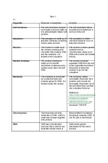
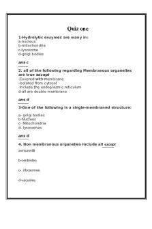
Cell biology lecture notes PDF

| Title | Cell biology lecture notes |
|---|---|
| Course | Biotechnology |
| Institution | SRM Institute of Science and Technology |
| Pages | 108 |
| File Size | 1.2 MB |
| File Type | |
| Total Downloads | 100 |
| Total Views | 261 |
Summary
Cell Biology Lecture Notes Introduction: A. Definition of a cell: fundamental structural and functional unit of all living organisms B. Characteristics of cells: 1) Contain highly organized molecular and biochemical systems and are used to store information 2) Use energy 3) Capable of movement 4) Se...
Description
Cell Biology Lecture Notes Introduction: A. Definition of a cell: fundamental structural and functional unit of all living organisms B. Characteristics of cells: 1) Contain highly organized molecular and biochemical systems and are used to store information 2) Use energy 3) Capable of movement 4) Sense environmental changes 5) Can duplicate (transfer genetic information to offspring) 6) Capable of self-regulation -Most cells are microscopic (invisible to the naked eye) and thus, a microscope is needed to view most cells. C) History: -Discovery of the cell followed by the development of the microscope A. 1665-Robert Hooke- observed cells from the fruiting bodies of fungi B. Anton van Leewenhoek- observed a variety of cells and called them "animalcules" C. 1830’s-Theodor Schwann and Matthias Schleiden developed the cell theory -Cell Theory states: 1. 2. 3.
All living organisms are composed of cells Cells are the functional units of living organisms Cells arise from preexisting cells via division
D) Louis Pasteur-developed the theory of spontaneous generation that is that cells could develop from non-living matter -Also worked on problem associated with the fermentation of French wine -1857-developed a partial sterilization process called pasteurization- involves heating at a moderate temperatures to reduce the number of living microorganisms E) 1865-Mendel-demonstrated that cellular traits (phenotypes) were inherited - Seed shape and color in garden peas - Named "Father of Genetics"
F) 1871-Johan Freidrick Miescher-isolated nucleic acids from cells "nuclein"
G) 1889-R.Altman-purified nucleic acids
H) 1944-Oswald Avery, Colin MacLeod, and MacLyn McCarty-Demonstrated that DNA was the heredity molecule -DNA could transform bacterial cells I) 1952-Alfred Hershey and Martha Chase-also demonstrated that DNA was the heredity molecule -Radioactive DNA from a virus was able to infect and transform bacterial cells J) 1953-James Watson and Francis Crick-developed the 3-D structure of DNA
K) 1958-Mattew Meselson and Frank Stahl-demonstrated that DNA replicated by a semi conservative method L) 1961-Brenner, Jacob, Meselson-discovered RNA M) 1966-Nirenberg and Khorana-elucidated the chemical nature of the genetic code N) 1972-1973-Berg, Boyer, and Cohen- discovered gene cloning O) 1975-Gilbert and Sanger-developed chemical techniques to rapidly sequence DNA Cell Structure: I. II. III.
Most cells are microscopic and cannot be seen by the naked eye. Microscopes were developed to visualize cells. Resolution is the minimum distance where 2 objects can be visually separated
-Unresolved -Partially resolved -Resolved -Depends on: a. b. c.
Wavelength of light Refractive index of the medium Of the light
-The naked eye can resolve two separate objects separated by 200 um Metric system: -1 meter = 3.3 feet, 1 km = 103 m, 1cm = 10-2 m, 1mm = 10-3 m, 1um = 10-6 m, 1nm = 10-9 m, 1 A = 10-10 m, 1pm = 10-12 m
IV. Light microscope: -Can resolve two objects 100-200 nm apart (including cells and large sub cellular organelles) -Uses different light sources and patterns of image formation a. b.
Bright field d) differential interference Dark field e) fluorescence c) phase contrast
V. Electron Microscope: -Uses a beam of electrons (e-) rather than light as an illumination source A. Transmission Electron Microscope (TEM) -Electrons forming the image focused through the specimen -Short wavelength of e- beam improves the resolution of TEM to 5 A (.5nm) -Can resolve small sub cellular organelles and large proteins B. Scanning Electron Microscope (SEM) -Used to examine surfaces of cells or isolated cellular structures -e- beam "scans" the specimen -Resolution 5 to 10 nm Prokaryotic Cells- small and primitive bacteria and blue-green algae (cyanobacteria) Greek: Pro=before karyon=nucleus -Lacks specialized internal membrane-bound compartments known as organelles -Cell membrane- functions in transport, the movement of substances in and out of the cell, and in energy production (breakdown of large molecules, photosynthesis) -Cell wall- gives structural strength (rigidity) to the cell -Capsule- jelly-like substance which protects the cell wall from environmental damage -Nucleiod- contains a single circular molecule of DNA (stores genetic information) -Cytoplasm- region surrounding the nucleiod and within the cell membrane -Contains ribosomes and RNA (site of protein synthesis) -Vacuole (vesicles)(blue-green algae)-site of photosynthesis (storage) -Flagellum- protein fiber the functions in movement
Eukaryotic Cell- (eu=true karyon=nucleus) 1. 2. 3.
Possesses a complex membrane system Has a true nucleus Distinct membrane-bound intracellular compartments called organelles -Nucleus- dark-staining body within the cell by enclosed an intracellular membrane called the nuclear envelope
-Nuclear envelope contains pores, which are filled with a ring of proteins called annulus -Contains DNA in the form of chromatin fibers -DNA is linear (linear DNA + proteins = chromosome) -Nucleolus- a cell organelle in the nucleus that disappears during part of cell division. Contains rRNA genes -Nucleus also contains RNA (mRNA, rRNA, and tRNA) -Transcription- conversion of genetic information from DNA to RNA occurs in the nucleus -DNA replication-duplication of genetic material -Cytoplasm: major portion of the protoplasmic substance within the cell membrane a.
Ribosomes-a cytoplasmic particle that contains RNA and protein and is involved in the process of protein synthesis
-Translocation-process which takes place in the cytoplasm and converts genetic information in RNA into proteins -Ribosomes can either be freely suspended in the cytoplasm or attached to intracellular membranes a.
Endoplasmic reticulum (ER)- a network of intracellular membranes where secreting proteins are synthesized -Rough ER- the ER + ribosom es -Smooth ER- the ER without ribosom es Function
s in the breakdo wn of fats attached to the rough ER in the Golgi complex a.
Golgi apparatus-a membranous organelle that packages and sorts newly synthesized secretory proteins
a.
Lysosome- organelle which contains digestive enzymes e. Mitochondrion-semiautonomous eukaryotic cell organelle -Site of respiration -Consists of an outer membrane and a convoluted inner membrane -Site of ATP production within the cell
a. b.
Microbody-organelle within a cell containing specialized enzymes whose functions involve hydrogen peroxide (peroxisome) Microtubules-composed of tubulin
h. Microfilaments-composed of actin -Both (g and h) are involved in cellular movement a. b.
Intercellular-includes flagella and cilia Intracellular- cytoplasmic streaming
Plant cell organelles: -Chloroplast- involved in photosynthesis -Central vacuole- provides support to the plant via osmotic pressure -Cell wall- composed of cellulose, which provides extra strength and rigidity i. Specialized protozoan cell organelle: -Contractile vacuole- used to maintain proper osmotic pressure and secretes waste and excess H2O -Two types of nuclei 1) Macronucleus- involved in asexual reproduction
2) Micronucleus- involved in sexual reproduction Chemical Bonds: I. 4 types of molecules make up cells: 1) Carbohydrates 2) Lipids 3) Proteins 4) Nucleic acids II. Biological macromolecules are held together by several different types of bonds: 1) Ionic bond-a transfer of electrons 2) Covalent bond-the sharing of electrons 3) H-bonds-weak attraction when H+ serves as a bridge between 2 electronegative atoms by a covalent bond and electrostatic attraction 4) Nonpolar associations-hydrophobic vs. Hydrophilic 5) Van der Waals-a momentary dipole that will affect the electron distribution of neighboring molecules Acids and Bases: Lewis definition: 1. 2. 3. 4. 5. 6. 7.
Acid-a substance that can take up an electron pair to form a covalent bond Base-a substance that can donate an electron pair to form a covalent bond H2O dissociates into H+ ions and OH[H+] + [OH-] = 1x10-14 moles/liter (M) pH = -log10 [H+] Acid pH is from 0 to 7 Base pH is from 7 to 14
-Condensation reaction-when two molecules are combined into one molecule with the release of one water molecule - A + B == C + H2O Ex: 2 amino acids are joined together to form a dipeptide molecule -Hydrolysis reaction-when one molecule is broken into two molecules with the addition of water molecule - C + H2O == A + B Ex: disaccharide maltose + water == 2 glucose molecules
Reactive Organic Molecules:
1. Hydroxyl group - strongly polar and highly reactive 2. Carbonyl group - weakly polar and highly reactive 3. Aldehyde 4. Ketone 5. Carboxyl group - strongly polar and acts as an acid 6. Amino group - polar and acts as a base 7. Phosphate group - acidic and polar 8. Sulfhydral group - readily oxidized -Two sulfhydral groups can bond together to form a disulfide bond Carbohydrates: A. Function: 1. 2. 3.
Store energy (starches in plants / glycogen in animals) Provides rigidity to plant cells (cellulose) Involved in cell-cell communication (glycoproteins)
B. Structure -Carbohydrates have a characteristic content of C, H, O atoms in the ratio of 1C:2H:1O 1) Monosaccharide is the subunit of a carbohydrate 2) Disaccharide contains 2 monosaccharide subunits 3) Oligosaccharide contains 2-10 monosaccharide subunits 4) Polysaccharide contains >10 monosaccharide subunits C. Most carbohydrate subunits contains 3 carbons (triose), 5 carbons (pentose), or 6 carbons (hexose) glucose - glucose - glucose D. 2 common monosaccharides are: fructose and galactose
E. Disaccharide = 2 monosaccharide subunits linked together
-Ex: maltose = 2 glucose molecules linked together -Glycosidic bond = is the bond between 2 carbohydrate subunits formed by the eliminated of water F.
Examples of Polysaccharides:
1) Cellulose- comprises plant cell walls; molecule composed of repeating B- glucose units (monomers) held together by B 1=4 linkages 2) Starch- (primary storage compound in plants) is a macromolecule composed of repeating - glucose units held together by 1=4 linkages 3) Glycogen- (primary storage compound in animals) is a branched macromolecule composed of repeating 1=4 and 1=6 glycosidic linkages Sucrose:
Lactose:
-Most monosaccharides can exist in alternative forms when molecules, which are attached to the carbon chain, can be oriented in different positions -Stereoisomers - two molecules, which have the same molecular formula and the same chemical formula and physical properties, but are different in the spatial arrangement of atoms -Most carbohydrates exist in D and L forms Lipids:
A. Definition- fats or fat-like substances that are insoluble in water and soluble in nonpolar solvents like acetone, ether, chloroform and benzene. B. Function: 1) Primary component of cell membranes 2) Store energy C. There are 3 types of lipids: neutral lipids, phospholipids, and steroids D. Neutral lipids (fats and oils) 1) Composed of fatty acids and glycerol (alcohol) 2) Fatty acid is a long, unbranched chain of carbon atoms attached by hydrogen and other groups and a terminal carboxyl group 3) CH3(CH2)nCOOH saturated fatty acid since the carbons have the maximum possible # of H atoms 4) CH3(CH2)nCH=CH(CH2)nCOOH unsaturated fatty acid because of the one double C-C bond. 5) Structure of neutral lipids
- Glycerol has 3 OH groups each of which is attached to a fatty acid E. Phospholipids-primary lipids in cell membranes -Most common phospholipid is a phosphoglyceride -2 fatty acids + glycerol + phosphate group -One end is hydrophobic; the other end is hydrophilic =called amphipathic (amphiphilic) -To satisfy these solubility properties, phospholipids arrange themselves into a lipid bilayer -The lipid bilayer is the basic arrangement of the cell membrane F. Steroids -Based on a framework of 4 carbon ring
-Sterols are the most abundant group of steroids
-A nonpolar side chain is attached at one end of the ring structure and a polar side group is attached to the opposite end of the ring structure -R’ is a polar unit and R is a nonpolar unit -The combination of polar and nonpolar side groups gives phospholipids dual solubility properties -An example of a sterol is cholesterol
-Cholesterol is an important component of the cell membrane in all animal cells -Cholesterol also can be deposited inside arteries causing blockage, which contributes to the disease arteriosclerosis (hardening of the arteries) -Hormones are steroids and they play major roles in cell regulation, cell metabolism, and cell growth I. Proteins carry out many cellular functions: 1) Provide cellular support (cytoskeleton: microtubules – tubulin; microfilaments - actin) 2) As enzymes, they catalyze cellular reactions 3) Stabilize and control gene activity via interactions with other proteins and nucleic acids (DNA or RNA) 4) Used in cell transport and cell recognition (cell membrane) 5) Involved in cell-cell transport via secretory proteins and hormones II. Proteins are composed of subunit structures called amino acids A. 20 major biological amino acids B. General structure of amino acid
C. 20 amino acids - commit to memory: 1) Structure 2) Name 3) 3 letter abbreviation 4) 1 letter abbreviation D. Amino acids can be linked together in chains of 2 or more units
-Peptide bond - is a bond in which the carboxyl group of one amino acid is joined to the amino group of a second amino acid via a condensation reaction -Peptide - is a chain composed of 2 or more amino acids and contains one or more peptide bonds -Ex: dipeptide = chain composed of 2 amino acids -Tripeptide = chain composed of 3 amino acids -Polypeptide is an amino acid chain composed of 3 or more amino acids -The amino acid sequence of a polypeptide chain is called the primary structure of a protein E. Secondary structure of proteins - is the conformation imposed on the polypeptide chain by hydrogen bonding between amino acids - There exists physical constraints on the rotation of the alpha carbon atoms that flank the peptide bond - It has been determined that there are only 2 or 3 stable arrangements of amino acids which conform to these restraints 1. 2. 3.
Alpha helix Beta strands (sheets) Random coil - These arrangements compose the secondary structure of a polypeptide chain
-Secondary structure - is the arrangement of alpha helices, beta sheets, and random coils in a polypeptide chain 1) Alpha helix - common structural motif of a polypeptide chain in which the linear sequence of amino acids folds into a right-handed helix -Helix is stabilized by internal hydrogen bonding between backbone atoms 2) Beta sheet - common structural motif of a polypeptide chain, which is composed of beta, strands that are oriented in an antiparallel fashion -Stabilized by internal hydrogen bonds -Beta strand - is an extended zigzag arrangement of amino acids in a polypeptide chain -Beta barrel - is a cylindrical arrangement of beta sheets -Example of a protein that is composed primarily of beta sheets is the silk protein secreted by silk worms (contributes to the high strength of silk fibers) 3) Random coil - an irregular configuration of amino acids within a polypeptide chain -Usually composed of proline - cannot fit into an alpha helix or beta sheet -Allows the protein to bend and flex
-Allows the protein to compact into its most stable energetic structure F) Tertiary structure of proteins -The three-dimensional arrangement of a polypeptide chain within a protein (monomeric protein) G) Quaternary structure of protein -Three-dimensional relationship between 2 or more polypeptide chains within a complex protein -Ex: coiled coil & triple helix -Dimer = 2 subunits -Homodimer = identical subunits -Heterodimer = distinct subunits -Multimeric protein - composed of 2 or more subunits (identical or distinct) Nucleic acids: I. Definition - a large, chain-like macromolecule containing phosphoric acid, sugar, and a nitrogenous base -2 examples are deoxyribonucleic acid (DNA) & ribonucleic acid (RNA) a. Sugar is 5-carbon sugar called a pentose ribose deoxyribose
b. Phosphoric acid is composed of one or more phosphate groups (PO4-) c. Nitrogenous base = 2 types purine pyrimidine
1. Two common purine bases, adenine and guanine 6-aminopurine (adenine) 2-amino-6-hydroxypurine (guanine)
2. Three common pyrimidine bases, cytosine, thymine, and uracil 4-amino-2-hydroxypyrimidine 2,4-dihydroxypyrimidine 2,4-dihydroxy-5-methyl pyrimidine (cytosine) (uracil) (thymine)
II. Nucleoside - macromolecule composed of a nitrogenous base joined to a pentose III. Nucleotide - is a macromolecule composed of a nitrogenous base, a pentose, and linked (esterified) to one or more phosphate groups -5’-ribo(deoxyribo) - nucleotide - Phosphate is linked to the 3’ OH of the pentose = 3’-ribo(deoxyribo) - nucleotide IV. Nomenclature of nucleosides: A-deoxyriboside = deoxyadenosine A-riboside = adenosine G-deoxyriboside = deoxyguanosine G-riboside = guanosine C-deoxyriboside = deoxycytidine C-riboside = cytidine T-deoxyriboside = deoxythymidine U-riboside = uridine V. Nomenclature of nucleotides in DNA base - deoxyribose - phosphate 5’-dAMP = deoxyadenosine-5’-PO4 5’-dGMP = deoxyguanosine-5’-PO4 5’-dTMP = deoxythymidine-5’-PO4
5’-dCMP = deoxycytidine-5’-PO4 VI. Nomenclature of nucleotides in RNA base - ribose - phosphate 5’-AMP = adenosine-5’- PO4 5’-GMP = guanosine-5’- PO4 5’-UMP = uridine-5’- PO4 5’-CMP = cytidine-5’- PO4 VII. Nomenclature of nucleoside triphosphates ATP = adenosine-5’-triphosphate dATP = deoxyadenosine-5’-triphosphate VIII. Nucleic acids RNA and DNA -Nucleotides held in chains by bridging a phosphate group that extends between the 5’-carbon of one sugar with the 3’-carbon of a second sugar (held together by a phosphodiester bond) -Produces a backbone chain of alternating sugar and PO4 groups -DNA exists in a double helix that contains 2 intertwined chains of nucleotides -RNA is single-stranded Enzymes: 1. Review thermodynamics -First and second law of thermodynamics -Reversible reactions -Coupling reactions -Standard free energy change 2. Definition of an enzyme - is a protein which increases the rate of a spontaneous reaction (catalyzes the reaction) a) Lowers the activation energy of the transition state b) Reaction would proceed without the enzyme c) Enzyme cannot make a reaction occur that would not proceed spontaneously without the enzyme
d) Enzymes do not alter the equilibrium of a reversible reaction e) Enzymes increase the rate at which a reaction reaches equilibrium Classification of enzymes: 1. Oxidoreductases - catalyzes a reaction in which electrons are removed from the substrate are donated directly to molecular oxygen -Catalyzes oxidation-reduction reactions -Act on alcohols, ketones, aldehydes, amines, etc. 2. Transferase - catalyze the transfer of functional groups -Sulfhydral, glycosyl, aldehyde, acyl, etc.
3. Hydrolases - catalyze hydrolysis reactions -Glycosidic bonds -Peptide bonds 4. Lyases - catalyze the addition of groups to double bonds - C=C, C=N, C=O -Ex: AP lyases involved in repairing DNA 5. Isomerases - catalyze an intramolecular rearrangement -Catalyzed isomerization reaction -Isomerization - rearrangement of atomic group within the same molecule without any loss or gain of atoms 6. Ligases - a group of enzymes that catalyze reactions in which a bond is formed between 2 substrate molecules using energy (ATP) obtained from the cleavage of a pyrophosphate bond -Ex: DNA ligase & RNA ligase
Characteristics of enzymatic proteins: -Enzymes combine briefly with reactants during an enzyme-catalyzed reaction (enzyme-substrate complex) -Enzymes are released unchanged after catalyzing the conversion of reactants to products
-Enzymes are specific in their activity; each enzyme catalyzes ...
Similar Free PDFs

Cell biology lecture notes
- 108 Pages

Cell Biology - Lecture notes 1
- 35 Pages

Cell biology revision notes
- 5 Pages

Cell Biology Lecture Notes - Test 4
- 17 Pages

Cell biology L2-12 notes
- 21 Pages

Cell biology summary notes website
- 33 Pages

Unit 1 Cell Biology - notes
- 5 Pages

Cell biology
- 3 Pages

Lecture notes, Cell Bio
- 3 Pages

CELL Cycle - Lecture Notes
- 3 Pages

Cell Biology Quiz CELL ORGANELLES
- 10 Pages

Cell Adhesion Lecture Notes
- 6 Pages
Popular Institutions
- Tinajero National High School - Annex
- Politeknik Caltex Riau
- Yokohama City University
- SGT University
- University of Al-Qadisiyah
- Divine Word College of Vigan
- Techniek College Rotterdam
- Universidade de Santiago
- Universiti Teknologi MARA Cawangan Johor Kampus Pasir Gudang
- Poltekkes Kemenkes Yogyakarta
- Baguio City National High School
- Colegio san marcos
- preparatoria uno
- Centro de Bachillerato Tecnológico Industrial y de Servicios No. 107
- Dalian Maritime University
- Quang Trung Secondary School
- Colegio Tecnológico en Informática
- Corporación Regional de Educación Superior
- Grupo CEDVA
- Dar Al Uloom University
- Centro de Estudios Preuniversitarios de la Universidad Nacional de Ingeniería
- 上智大学
- Aakash International School, Nuna Majara
- San Felipe Neri Catholic School
- Kang Chiao International School - New Taipei City
- Misamis Occidental National High School
- Institución Educativa Escuela Normal Juan Ladrilleros
- Kolehiyo ng Pantukan
- Batanes State College
- Instituto Continental
- Sekolah Menengah Kejuruan Kesehatan Kaltara (Tarakan)
- Colegio de La Inmaculada Concepcion - Cebu


