Chapter 6 - Lecture notes 6 PDF

| Title | Chapter 6 - Lecture notes 6 |
|---|---|
| Course | Exercise Physiology |
| Institution | University of Delaware |
| Pages | 11 |
| File Size | 157.4 KB |
| File Type | |
| Total Downloads | 38 |
| Total Views | 208 |
Summary
notes...
Description
Chapter 6: The Cardiovascular System and Its Control
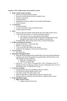
Major Cardiovascular Functions o Delivery of oxygen and other nutrients o Removal of carbon dioxide and other metabolic waste o Transport of hormones o Thermoregulation o Maintenance of acid-base balance and overall body fluid balance o Immune function 3 Components o A pump (the heart) o A system of channels or tubes (the blood vessels) o A fluid medium (the blood) Heart o Primary pump that circulates blood through the entire cardiovascular system o 2 Atria (receiving chambers) 2 Ventricles (pumping chambers) The valves are one-way valves that prevent backflow Left ventricle is most powerful b/c it generates enough blood to pump through the entire body so it has the thickness muscular wall o Pericardial fluid: reduces friction between the sac and the beating heart Position of the Heart in the Chest o Top part of the heart: 2nd intercostal space o Bottom: 5th intercostal space o 2/3 lies to the left of the middle of the sternum o Attached of the diaphragm, sternum and vertebral column Size of the Heart o Size of your fist Blood Flow Through the Heart o Right side of heart pumping deoxygenated blood to the lungs through pulmonary circulation and the left side pumping oxygenated blood to all other tissues in the body through systemic circulation Right side = Pulmonary Side Left side = Systemic Side o Into the IVC + SVC RA Tricuspid Valve RV Pulmonary Valve Pulmonary Trunk Pulmonary Arteries Lungs Pulmonary Veins LA Mitral Valve LV Aortic Valve Aorta Coronary Circulation o Coronary arteries lie on the epicardium (outer most surface) o Left anterior descending supplies anterior wall o Circumflex supplies later and posterior wall o Right coronary arteries supplies inferior (apical) or posterior wall o Posterior wall is the only part of the heart that has 2 different blood sources Myocardial Blood Supply o Right coronary artery Supplies right side of the heart
Divides into marginal, posterior interventricular o Left (main) coronary artery Supplies left side of the heart Divides into circumflex, anterior descending o Atherosclerosis (narrowing by the accumulation of plaque and inflammation) coronary artery disease The Anatomy of the Human Heart Myocardium- The Cardiac Muscle o Thickness varies according to the stress placed on chamber walls o Left ventricle is the most powerful of chambers and the largest chamber o With exercise training, the size of the left ventricle increases o Intercalated disks in the myocardium allow impulses to travel quickly in cardiac muscle and allow it to act as one large muscle fiber; all fibers in the heart contract together as one unit Mechanism of Cardiac Muscle Contraction o Contraction occurs by Ca-induced and Ca-released: Ca enter through DHPR receptor in T-tubules and triggers the RYR receptor Ca is then released from the SR Structural and Functional Characteristics of Skeletal and Cardiac Muscle o Skeletal: striated, voluntary Location: Named muscle attached to the skeleton and fascia of limbs, body wall and head/neck Appearance: large, long, unbranched, cylindrical gibers with transverse striations (stripes) arranged in parallel bundles, multiple peripherally located nuclei Type of Activity: Strong, quick intermittent (phasic) contraction above a baseline tonus, acts primarily to produce movement or resist gravity Simulation: Voluntary (or reflexive) by the somatic nervous system o Cardiac: striated, involuntary Location: Muscle of the heart (myocardium) and adjacent portions of the great vessels (aorta, vena cava) Appearance: branching and anastomosing shorter fibers with transverse striations (stripes) running parallel and connected end to end by complex junctions (intercalated disks): single, central nucleus Type of Activity: strong, quick, continuous rhythmic contraction, pumps blood for the heart Simulation: Involuntary, Intrinsically (myogenically) stimulated and propagated; rate and strength of contraction modified by the autonomic nervous system o Cardiac Cells- Conductivity Cardiac muscle has the unique ability to generate its own electrical signal (spontaneous rhythmicity) Conduction Velocity Varies in different Cardiac tissue AV node: 200 mm/s V. Muscle: 400 mm/s A. Muscle: 1000 mm/s
Perkinjie: 4000 mm/s o Intrinsic Conduction System of the Heart o Cardiac Conduction System Sinoatrial (SA) node: pacemaker of the heart Atrioventricular (AV) node: receives electrical impulse from SA node and tavels to AV bundle, impulse is delayed to allow blood from the atria to completely empty into the ventricles to maximize ventricular filling before the ventricles contract AV bundle (bundle of His): sends impulse along the ventricular septum to the respective branches and divides into terminal branches known as Purkinjie fibers Purkinjie fibers: transmit impulse through the ventricles SA node is connected to AV node by internodal pathways o Intrinsic Control: Automaticity of the Heart The heart has its own inherent rhythm dependent upon what part of the heart initiates the impulse In addition, the cardiac cells transmit electric impulses very rapidly due to their structure SA: 60-100 bpm AV: 40-60 bpm Ventricles: 15-40 bpm If SA node fails to fire, AV node takes over as pacemaker If AV fails to fire or impulse gets blocked, the ventricles and purkinjie fibers take over as pacemaker o Conduction System and ECG P Wave: Atrial depolarization and occurs when the electrical impulse travels from the SA node through the atria to the AV node QRS Complex: ventricular depolarization occurs as the impulse spreads from the AV bundle to the Purkinjie fibers and through the ventricles T Wave: represents ventricular repolarization o Extrinsic Control of the Heart ANS & Endocrine System Although the heart initiates its own electrical impulses, both the heart rate and force of contraction can be altered Parasympathetic Nervous System (PNS) acts through the vagus nerve, releasing Ach to decrease heart rate and force of cardiac contraction Vagus nerve has a depressant effect on the heart. It slows impulse generation and conduction and thus decreases the heart rate Hyperpolarization of the conduction fells Absence of vagal tone, HR= 100 beats/min Maximal vagal tone, HR= 20-30 beats/min Sympathetic Nervous System (SNS) increases rate of impulse generation and conduction speed, increasing heart rate and force of cardiac contraction Maximal sympathetic stimulation, HR= 250 beats/min Sympathetic control dominates during times of physical or emotional stress when the heart is greater than 100 beats/min
o
o
o
o
o o
When exercise begins, HR first increases due to withdrawal of vagal tone but then increases further to do sympathetic activation Endocrine System (hormones: Epinephrine and norephinephrine): released from the adrenal medulla as a result of sympathetic stimulation: increase heart rate and contractility Parts of the Cardiovascular System CR Center is located in the Medulla Oblongata Autonomic Nervous System Parasympathetic nerve fibers supply SA and AV nodes and the atrial muscle by means of the vagus nerves Acetylcholine: neurotransmitter released when PS nerves re stimulated, slows HR and conduction rate through AV node Sympathetic nerves supply specific areas of the conduction system, atrial muscle and ventricular muscle Norepinephrine and Epinephrine neurotransmitters acts on Beta 1 receptor in heart Increases HR, BP and force of contraction Relative Contribution of Sympathetic and Parasympathetic Nervous Systems to the Rise in heart Rate During Exercise When HR is under 100, HR increases by decreasing activity of the parasympathetic nervous system When HR is over 100, HR increases by increasing activity of the sympathetic nervous system Heart Rate and Endurance Training Resting HR in adults tend to be between 60 and 85 beats per min Extended endurance training can lower resting HR to 35 beats or lower Lower heart rate is thought to be due to increased parasympathetic stimulation Electrocardiogram (ECG) An ECG provides a graphical record of the electrical activity of the heart and can be used to aid clinical diagnoses Represents the events occurring in one cardiac cycle (contraction/relaxation) 1/3 of the cardiac cycle is systolic, 2/3 of the cardiac cycle is dystolic 12 Lead ECG Cardiac Anatomy, Conduction, and Control Key Points The atria receive blood from the veins; the ventricles eject blood from the heart The left ventricular myocardium is larger because it must produce more force than the other ventricles to pump blood to the systemic circulation Cardiac tissue is capable of spontaneous rhythmicity and has its own conduction system The SA node normally establishes heart rate
o
o
o
o
Heart rate and contractility can be altered by the PNS, SNS, and the endocrine system The ECG is a recording of the heart’s electrical activity Cardiac Arrhythmias Arrhythmia: irregular heart rhythm Bradycardia: resting HR below 60 bpm Tachycardia: resting HR above 100 bpm Bradycardia/Tachycardia may affect SBP: symptoms include fatigue, dizziness, lightheadness, fainting Premature ventricular contractions (PVCs): skipped or extra beats from impulses originating outside the SA node Ventricular tachycardia: 3 or more consecutive PVCs Ventricular fibrillation: depolarization (contraction) of the ventricular tissue is uncoordinated and can result in cardiac death Atrial Flutter: atria depolarize at rates of 200 to 400 beats/min Atrial Fibrillation: atria depolarize in a rapid and uncoordinated manner Endurance Training vs. Pathological Bradycardia The decrease in resting HR that occurs as an adaptation to endurance training is different from pathological bradycardia, an abnormal disturbance in resting HR Cardiac Cycle Defined as the mechanical and electrical events that occur during one heart beat (systole to systole) Diastole is the relaxation phase during which the chamber fill with blood (T wave to QRS) Diastole 62% of the cardiac cycle. At HR of 74, entire cardiac cycle is .81 sec. The heart is filling (diastole) for .5 sec. Diastole is almost 2 times longer than systole Systole is the contraction phase during which the chambers expel blood (QRS to T wave) 38% of a cardiac cycle During ventricular diastole, the pressure inside the ventricles is low, allowing the ventricles to passively fill with blood. As the ventricles contract, pressure inside the ventricles increases, which forces the atrioventricular valves (tricuspid and mitral) to close and prevent backflow. Then when ventricular pressure exceeds the pressure in pulmonary artery and aorta, the pulmonary and aortic valves open, allowing blood to flow into the pulmonary and systemic circulations. After ventricular contraction, pressure inside the ventricles decrease and the pulmonary and aortic valves close. Stroke Volume and Cardiac Output Stroke Volume (SV) Volume of blood pumped per contraction End-Diastolic Volume (EDV): volume of blood in ventricle just before contraction (100 ml)
End-Systolic Volume (ESV): volume of blood in ventricle just after contraction (40 ml) SV= EDV - ESV (100ml-40ml = 60ml) SV= 60 ml/beat, SV varies between 60-80 ml/beat in avg person Cardiac Output (Q) Total volume of blood pumped by the ventricle per minute Q= HR x SV Q= 83 beats/min x 60 ml/beat = 5000 ml/min or 5 L/min o Ejection Fraction (EF) Fraction of blood pumped out of the left ventricle with each beat EF = SV/EDV, expressed as a percentage Averages 60% at rest in healthy individuals o Considerably lower in CHD patients 60% of blood in ventricle is pumped out when the heart contracts o Calculation of Stroke Volume, Ejection Fraction, and Cardiac Output Stroke Volume units = ml/beat Ejection fraction is a percentage Cardiac Output is ml/min or L/min o The Vascular System Arteries: largest (aorta), most muscular, most elastic, carry blood AWAY from the heart arterioles Arterioles: site of greatest control by SNS: from arterioles blood enters capillaries Capillaries: most narrow/simple, walls 1 cell thick, gas exchange occurs between tissues and capillaries, blood leaves capillaries to begin the return trip to the heart in venules Venules: vessels that start the return back to heart that lead into veins, eventually become larger veins Veins: IVC, SVC (largest veins) transport blood back to right atrium 85% muscles and 15% organs because of arterioles (they direct blood) 65% of your blood at one point is usually in your veins Blood Pressure o Pressure exerted by the blood on the arterial walls o Systolic blood pressure (SBP) is the highest pressure within the vascular system generated during cardiac contraction o Diastolic blood pressure (DBP) is the lowest pressure within the vascular system when the heart is relaxed o Mean arterial pressure (MAP) is the average pressure exerted as the blood moves through the arteries MAP = 2/3 DBP + 1/3 SBP (.67*.80 + .33*120) = 93mmHg MAP: DPB + [.33 (SBP – DBP)] MAP = 80 + [.33 (120-80)] = 93 mmHG o Optimal is now considered 110/70 General Hemodynamics o Cardiovascular system is a continuous, closed-loop
o Blood flow from a region within the vessel of high pressure to a region within the vessel with lower pressure (pressure gradient) No pressure = No blood flow o In aorta pressure is 100mmHG, in RA its close to 0 mmHg o Pressure gradient across the entire cardiovascular system = 100 mmHg o Blood vessels and the blood itself provide resistance to blood flow. Resistance determined by properties of the vessels and blood o Resistance to blood flow = [ηL /r^4] η = viscosity of the blood (thickness) L = length of vessel r^4= radius of vessel to the 4th power: MOST IMPORTANT VARIABLE Viscosity of blood and length of vessel doesn’t change too much but a tiny change in the radius of the vessel produces a huge change in the resistance to blood flow Changing Blood Flow o Blood Flow = Δpressure / resistance o Blood flow can change by either changing pressure or resistance or a combination of the two o Changing resistance has a larger effect on blood flow because of the fourth power mathematical relationship between vascular resistance and vessel radius o Blood flow regulation largely accomplished by small changes in vessel radius Vasoconstriction: radius of the vessel decreases, decreasing blood flow Vasodilation: radius of the vessel increases, increasing blood flow Arterioles: key role, most resistance to blood flow occurs here Pressure Changes Across the Systemic Circulation o Arterioles: responsible for 70-80% of decrease in mean arterial pressure o Small changes in arteriole diameter cause large changes in blood flow Distribution of Cardiac Output at rest and During Heavy Exercise o At rest, liver and kidneys combine to receive approximately half of the cardiac output, while resting skeletal muscles receive only about 15% to 20% o During exercise, contracting muscles may receive up to 80% of the blood flow and flow to the liver/kidneys decreases o During digestion, digestive system receives more of the available cardiac output o During heat, skin blood flow increases o Cardiovascular system responds accordingly to redistribute blood flow, this is controlled by the sympathetic nervous system, primarily by increasing or decreasing the diameter of the arterioles providing blood flow to the given tissue/organ Intrinsic Control of Blood Flow o Ability of local tissues to vasodilate/constrict to stimuli increased local blood flow o 3 Types of Intrinsic Control of blood flow
Metabolic Regulation: Metabolic factors local (changes in chemical components): increased oxygen demand: strongest stimulus (biggest factor) Increases in metabolic by-products (↑CO2, ↑LA ↑H+ ↓pH ↑°F ↑K+) (arterioles sensitive to changes; vasodilate to allow more blood flow; most sensitive to ↑ O2 need) Inflammatory chemicals Endothelium-Mediated Vasodilation: substances can be produced within the endothelium of arterioles that can initiate vasodilation in the vascular smooth muscle of those arterioles Nitric Oxide (vasodilation) Prostaglandins (vasodilation) Endothelium-derived hyperpolarization factors (EDHF) o Myogenic responses: pressure changes within the vessels themselves can also cause vasodilation/vasoconstriction The vascular smooth muscle contracts in response to an increase in pressure across the vessel wall and relaxes in response to a decrease in pressure across the vessel wall Intrinsic Control of Blood Flow o Arterioles: primary site for Control of Distribution of Blood Flow (vasodilate or vasoconstrict in response to the immediate environment) o Smooth muscle cells contract when pressure increases across vessel wall o Nitric Oxide, prostaglandins, endothelium-derived hyperpolarizing factor (EDHF) Extrinsic Neural Control o Accomplished by the sympathetic nervous system through vasoconstriction o SNS innervates smooth muscle cells (SMC) of the arteries and arterioles (most regulated site) o At rest, vessels are moderately constricted, which maintains BP; called vasomotor tone o When SMC are stimulated it causes vasoconstriction at the specific site and redistributed elsewhere Arteries have much more SMC innervated by SNS Extrinsic Control of BP o Blood pressure is maintained and controlled by the sympathetic nervous system o Normal condition vasomotor control o ↑SNS stim ↑vasoconstriction o ↓ SNS stim below level for vasomotor control vasodilation Integrative Control of Blood Pressure o Receptors that modify blood pressure controlled through the cardiovascular control centers (all 3 of these things influence what your BP is) Baroreceptors: stretch receptors in the aortic arch and carotid arteries that are sensitive to changes in blood pressure (↑BP ↑ stretchCV control center in brain ↑vagal ↓HR ↓SNS↓BP) Chemoreceptors: chemical receptors that relay information about the chemical environment, sensitive to oxygen, lactic acid
Mechanoreceptors: receptors that sense changes in muscle length and tension BP Classification System SBP DBP Normal...
Similar Free PDFs

Chapter 6 - Lecture notes 6
- 5 Pages

Chapter 6 - Lecture notes 6
- 6 Pages

Chapter 6 - Lecture notes 6
- 9 Pages

Chapter 6 - Lecture notes 6
- 11 Pages

Chapter 6 - Lecture notes 6
- 4 Pages

Chapter 6 - Lecture notes 6
- 3 Pages

Chapter 6 - Lecture notes 6
- 24 Pages

Chapter 6 lecture notes
- 1 Pages

Chapter 6 - Lecture Notes
- 16 Pages

COM Chapter 6 - Lecture notes 6
- 4 Pages

Chapter 6 slides - Lecture notes 6
- 49 Pages

MGMT - Chapter 6 - Lecture notes 6
- 14 Pages
Popular Institutions
- Tinajero National High School - Annex
- Politeknik Caltex Riau
- Yokohama City University
- SGT University
- University of Al-Qadisiyah
- Divine Word College of Vigan
- Techniek College Rotterdam
- Universidade de Santiago
- Universiti Teknologi MARA Cawangan Johor Kampus Pasir Gudang
- Poltekkes Kemenkes Yogyakarta
- Baguio City National High School
- Colegio san marcos
- preparatoria uno
- Centro de Bachillerato Tecnológico Industrial y de Servicios No. 107
- Dalian Maritime University
- Quang Trung Secondary School
- Colegio Tecnológico en Informática
- Corporación Regional de Educación Superior
- Grupo CEDVA
- Dar Al Uloom University
- Centro de Estudios Preuniversitarios de la Universidad Nacional de Ingeniería
- 上智大学
- Aakash International School, Nuna Majara
- San Felipe Neri Catholic School
- Kang Chiao International School - New Taipei City
- Misamis Occidental National High School
- Institución Educativa Escuela Normal Juan Ladrilleros
- Kolehiyo ng Pantukan
- Batanes State College
- Instituto Continental
- Sekolah Menengah Kejuruan Kesehatan Kaltara (Tarakan)
- Colegio de La Inmaculada Concepcion - Cebu


