PAR211 ACS - The clinical manifestations of ACS and how to treat as per QAS guidelines PDF

| Title | PAR211 ACS - The clinical manifestations of ACS and how to treat as per QAS guidelines |
|---|---|
| Author | Jordanna Hansen |
| Course | Cardiac and Respiratory Emergencies |
| Institution | University of the Sunshine Coast |
| Pages | 5 |
| File Size | 55.3 KB |
| File Type | |
| Total Downloads | 18 |
| Total Views | 150 |
Summary
The clinical manifestations of ACS and how to treat as per QAS guidelines...
Description
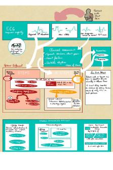
ACS Differential Diagnoses: Anxiety disorders Asthma Pericarditis Cardiomyopathies Stable angina Reflux Next Steps I would check the scene for danger, before introducing myself and my partner and asking why the patient has called for an ambulance. In the first 30 seconds on scene, I would conduct a primary survey and assess the patient’s airway and breathing condition before assessing if the patient is tripoding, clutching their chest, is pale, cyanosed or diaphoretic. This can indicate ACS. I would assess patient for signs of haemorrhage or spinal injury, before checking their pulse and assessing GCS. I would ask if their colouring and perspiration is normal for them. I would then conduct a SITREP and have my partner take vitals in this order: 1. Spo2 & pulse & respiratory rate/rhythm/strength 2. BP 3. ECG 4. Temperature 5. BGL While my partner is taking vitals, I would begin taking the history of the patient using SAMPLE and SOCRATES for the pain assessment. Based on their symptoms, it would be important to ask about a history of cardiac illness such as heart attacks or angina and establish if they are taking any medications for any possible illnesses. It would also be important to ask about events leading up to the call, as things like exercise could exacerbate cardiovascular illnesses (angina). I would next conduct a perfusion status assessment (BP, pulse, skin, cap refill) and a respiratory status assessment (CAPERRSSS). Questions: Cardiac causes: I would ask the patient if they have a past history of cardiovascular illness such as hyperlipidaemia/hypertension or diabetes mellitus. Family history of cardiovascular illness can be a risk factor as can a history of heart attacks or angina. I would also ask if they have previously been treated for cardiovascular illness (possible PCI) and about possible medications that have been prescribed, such as beta blockers as this can indicate hypertension or ectopic beats which is a risk factor for an acute cardiovascular illness. I would also ask about the onset, if it was acute or if it happens regularly and is relieved by rest (this can indicate stable angina). If they do have stable angina and it is now not relieved by rest, it may have become unstable. I would also
ask about their diet and exercise. Unhealthy habits such as a poor diet or regular smoking is a risk factor for ACS. Respiratory causes: I would ask the patient about their medical history. Have they been diagnosed with a respiratory illness or recently been sick with a cough or sore throat? I would also ask how long they had been feeling ill, if it is acute and suddenly came on or if it has been like this for a while and is exacerbated by something like exercise. I would ask if they are on any medications, such as a bronchodilator as this can indicate a respiratory illness such as asthma. I ask about their family history, as family history of respiratory illness can be a risk factor. I would also need to know social history, specifically their smoking history as this could indicate COPD (emphysema). Vitals I would first assess oxygen saturations. With any type of dyspnoea causing illness, I would be concerned about the risk of hypoxaemia because oxygen delivery to cells is compromised. If the patient has no history of respiratory illness, optimum saturation is 94-98%. If the patient has a history of COPD, saturations may normally be 88-92%. I would then assess respiratory rate. I would be looking for a normal rate of 12-20bpm. During a cardiac illness, patients are often tachypnoeic at rest which indicates that not enough oxygen is making it to the blood stream. I would next assess pulse and blood pressure to find out if the patient is hemodynamically stable, before taking a 12 lead ECG. This will show possible dysrhythmias or a cardiac cause of their symptoms. I would next take the patient’s temperature. During the respiratory assessment, I would listen to the patient’s chest. I would be listening for normal breath sounds or abnormal breath sounds, such as a wheeze or crackles. Diagnosis Based on the clinical manifestations and history, the patient is suffering from ACS. These conditions relate to myocardial ischemia and include STEMI, NSTEMI and unstable angina. Ischemia is caused by atherosclerosis, which is impaired blood flow due to the build-up of plaque in the arteries from modifiable (diet, exercise, smoking) and non-modifiable (age, family history) risk factors. Stable plaque will obstruct blood flow (angina), while unstable plaque can crack and leak its contents into the blood. Platelets congregate to seal the gap, which causes a thrombus, either partially blocking the artery, with surrounding ischemic or injured tissue (NSTEMI) or completely blocking the artery (STEMI). Clinical manifestations include prolonged cardiac pain, anxiety (impending doom), nausea/vomiting, dyspnoea, syncope and impaired myocardial function. Diagnosis is based on a 12 lead ECG. Unstable angina will not cause biomarker release and have possible ST-segment depression. NSTEMI will cause release of troponin and have no q wave. STEMI involves the full thickness of the myocardium and cause ST-segment
elevation, T wave changes and pathologic Q wave (25% of the depth of following R wave). Types: Changes in I and aVL indicate a high lateral MI (left circumflex) Changes in II,III & aVF indicate inferior wall MI & possible right ventricular MI (right coronary artery) Changes in V1 &V2 indicate septal wall MI (left anterior descending) Changes in V3&V4 indicate anterior wall MI (left anterior descending artery) Changes in V1-V3 &V7-V8 indicate Posterior wall MI (right coronary artery, left circumflex). Based on the clinical manifestations and ECG, the patient is suffering from a… Management The first step is assessing the patient condition through ECG within 10 minutes. If the patient has unstable angina or a NSTEMI I would consider oxygen therapy, GTN for unstable angina, aspirin, fentanyl for the pain and ondansetron for vomiting. I would call for CCP backup and transport to hospital. If the patient was presenting with a STEMI, I would do the same with urgency to make it to hospital within 1 hour for PCI and ensuring CCP assistance. PCI is Percutaneous Coronary Intervention. It involves getting a cardio angiogram to assess the blockage and inserting a wire with a balloon to place a stent, which restores blood flow. If I was more than an hour from hospital, I would consider fibrinolysis administration of tenecteplase, enoxaparin and clopidogrel. This would dissolve the thrombus, causing the blockage and restore blood flow. PCI is the preferred method of treatment but only if the patient is within one hour of a facility, has a GCS of 15 and ST elevation greater than 1mm in 2 contiguous limb leads or 2mm in 2 chest leads. Contraindications include history of serious systemic disease, suspected aortic dissection, elderly patient requiring assistance with ADLs, MI in an acute trauma. If conditions are not met, fibrinolysis should be considered. There is a 26-item checklist including contraindications such as pregnancy, internal bleeding, intracranial haemorrhage, left BBB, allergy, stoke, etc. This is because there are complications such as life-threatening stroke, haemorrhage or failure to achieve reperfusion. If the patient is approved for fibrinolysis their consent should be obtained, tenecteplase (weight calculated IV) should be administered then enoxaparin (30mg IV), then clopidogrel (300mg oral), then enoxaparin (1mg/kg, max dose 100mg). The patient should be then transported to hospital. Medications I would consider oxygen therapy when the patient’s oxygen saturation is less than 94% for a patient without COPD and less than 88% with COPD. Oxygen (gas) can be administered with a nebuliser mask running at 2-6L/min.
GTN is a vasodilator used to decrease preload and afterload. It is indicated for suspected ACS with pain, APO, autonomic dysreflexia and Irukandji. Its syndrome. Presentation is a sublingual spray (400mcg dose) or a 50mg/10ml ampule. I would administer 400mg (spray) at 5-minute intervals between BP checks. There is no max dose. Contraindications are BP under 100mmhg, heart rate under 50bpm or over 150bpm, allergy, CVA, head trauma or use of erectile dysfunction drug in last 24 hours. Desired response is to reduce pain/increase cardiac output Aspirin is an antiplatelet which inhibits platelet aggregation. It is indicated for suspected ACS or APO. Presentation is a 300mg tablet. I would administer 300mg. Contraindications include patients under 18, peptic ulcers, chest pain with overdose, bleeding or clotting disorders, GI bleeding or allergy. Desired response is to stop further clot formation. Ondansetron is an anti-emetic. It is indicated for significant nausea or vomiting. Presentation is a 4mg tablet or 4mg/2ml ampule. I would administer 4-8mg IV (max dose 8mg). Contraindications include allergy, long QT syndrome, apomorphine use, patients under 3. Desired response is to stop nausea/vomiting. Fentanyl is a narcotic analgesic to treat pain. It is indicated for sedation, autonomic dysreflexia, intubation and significant pain. Presentation is 100mg/2ml ampule. I would administer 25mcg IV every 5 minutes up to 100mg. Contraindications include allergy. Desired response is a decrease in pain. Tenecteplase is a fibrinolytic used to dissolve thrombi. It is indicated for STEMI patients who meet criteria. Presentation is 50mg powered and solvent graduated syringe. I would administer between 30mg and 50mg depending on weight. Contraindications include patients under 18 and over 75, uncontrolled hypertension, allergy, left BBB and history of thrombocytopenia. Desired outcome is dissolving the thrombus. Enoxaparin is anticoagulant used with tenecteplase to dissolve thrombi. Indicated for STEMI patients. Presentation is prefilled syringe 60ml/0.6ml or 100mg/1ml. I would administer 30mg IV. Contraindications include allergy or patients who don’t meet fibrinolysis checklist. Desired outcome is dissolving the thrombus. Clopidogrel is an antiplatelet used with tenecteplase and enoxaparin to dissolve thrombi. It is indicated for STEMI patients receiving fibrinolysis. Presentation is a 75mg tablet. I would administer 300mg with water. Contraindications include allergy, current clopidogrel or ticagrelor therapy, patients under 18, active bleeding or intracranial haemorrhage. Desired outcome is dissolving the thrombus....
Similar Free PDFs

ACS - ACS Infographic
- 2 Pages

ACS - Summary of lecture
- 8 Pages

ACUTE CORONARY SYNDROME (ACS
- 47 Pages

ACS+Study+Guide+Questions
- 1 Pages

Acs - ergkjeflwejfl, wjfl owfk
- 50 Pages

Laporan PKL ACS Jogja
- 50 Pages

ACS practice exam
- 18 Pages

ACS 3909 Assignment 2
- 2 Pages

ACS Exam practice
- 18 Pages

ACS 1803 - Lecture 5
- 2 Pages
Popular Institutions
- Tinajero National High School - Annex
- Politeknik Caltex Riau
- Yokohama City University
- SGT University
- University of Al-Qadisiyah
- Divine Word College of Vigan
- Techniek College Rotterdam
- Universidade de Santiago
- Universiti Teknologi MARA Cawangan Johor Kampus Pasir Gudang
- Poltekkes Kemenkes Yogyakarta
- Baguio City National High School
- Colegio san marcos
- preparatoria uno
- Centro de Bachillerato Tecnológico Industrial y de Servicios No. 107
- Dalian Maritime University
- Quang Trung Secondary School
- Colegio Tecnológico en Informática
- Corporación Regional de Educación Superior
- Grupo CEDVA
- Dar Al Uloom University
- Centro de Estudios Preuniversitarios de la Universidad Nacional de Ingeniería
- 上智大学
- Aakash International School, Nuna Majara
- San Felipe Neri Catholic School
- Kang Chiao International School - New Taipei City
- Misamis Occidental National High School
- Institución Educativa Escuela Normal Juan Ladrilleros
- Kolehiyo ng Pantukan
- Batanes State College
- Instituto Continental
- Sekolah Menengah Kejuruan Kesehatan Kaltara (Tarakan)
- Colegio de La Inmaculada Concepcion - Cebu


