Sensory Association Cortex PDF

| Title | Sensory Association Cortex |
|---|---|
| Course | Neurobiology of Disease |
| Institution | University of Texas at Austin |
| Pages | 3 |
| File Size | 40.4 KB |
| File Type | |
| Total Downloads | 46 |
| Total Views | 142 |
Summary
lecture notes with text book notes...
Description
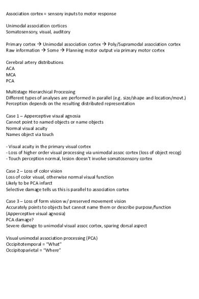
Association cortex = sensory inputs to motor response Unimodal association cortices Somatosensory, visual, auditory Primary cortex Unimodal association cortex Poly/Supramodal association cortex Raw information Some Planning motor output via primary motor cortex Cerebral artery distributions ACA MCA PCA Multistage Hierarchical Processing Different types of analyses are performed in parallel (e.g. size/shape and location/movt.) Perception depends on the resulting distributed representation Case 1 – Apperceptive visual agnosia Cannot point to named objects or name objects Normal visual acuity Names object via touch - Visual acuity in the primary visual cortex - Loss of higher order visual processing via unimodal assoc cortex (loss of object recog) - Touch perception normal, lesion doesn’t involve somatosensory cortex Case 2 – Loss of color vision Loss of color visual, otherwise normal visual function Likely to be PCA infarct Selective damage tells us this is parallel to association cortex Case 3 – Loss of form vision w/ preserved movement vision Accurately points to objects but cannot name them or describe purpose/function (Apperceptive visual agnosia) PCA damage? Severe damage to unimodal visual assoc cortex, sparing dorsal aspect Visual unimodal association processing (PCA) Occipitotemporal = “What” Occipitoparietal = “Where”
Language is localized to the supramodal cortex in the perisylvian region of L hemisphere Phoneme is a fundamental symbolic unit Broca’s area = motor / speech output = area 44/45 Wernicke’s area = sensory / language comprehension = area 22 Arcuate fasciculus links Broca and Wernicke’s areas (bidirectionally) AF is a part of the superior longitudinal fasciculus Case 4 – Broca’s aphasia Left hemisphere stroke that damages frontal lobe including Broca’s and (pre-)motor cortex Unable to produce much language, rather produces labored and slurred speech (dysarthric) Able to follow verbal and written instructions - Comprehension is intact - Repetition is impaired - Cortex for oral motor representation of phonemes damaged = dysarthric speech - Comprehension is intact bc Wernicke’s area is still intact - Brocas is actually located in the insula Case 5 – Conduction aphasia Infarct to area 22 Good comprehension of written and spoken language, decent speech Spontaneous language full of repeats and phonemic substitutions - Non-dysarthric speech, fluent - Phonemic paraphasias (substitutions) - intact comprehension - Impaired repetition - entire perisylvian language cortex used to convert words into speech, both areas intact Case 6 – Transcortical sensory aphasia - non-dysarthric speech - difficulty finding words - semantic paraphasic errors (uses wrong words) - impaired comprehension - repetition is unimpaired (because Broca’s/Wernicke’s are spared) - isolation of perisylvian cortex = generation and comprehension of speech impaired
Wernicke’s aphasia = transcortical sensory aphasia + conduction aphasia Fluent, non-dysarthric speech Semantic paraphasic errors Impaired comprehension Impaired repetition Apraxia = inability to carry out specific actions on command, eg use tools. Ventral visual association cortex (“what”) = used to recognize tool Left perisylvian cortex = used to name the tool Dorsal visual association cortex (“where”) = mind’s eye image of tool movement Premotor cortex = mind’s hand and arm of using the tool (this almost always opposite in terms of hemisphere to dominant hand) Ideomotor apraxia...
Similar Free PDFs

Sensory Association Cortex
- 3 Pages

Motorische cortex
- 3 Pages

Cortex Quercus
- 2 Pages

Cortex Frangulae
- 2 Pages

LAPORAN AKHIR FARMAKOGNOSI(Cortex)
- 15 Pages

C2 - Le cortex cérébelleux
- 4 Pages

Introduction to Sensory Systems
- 4 Pages

Word Association
- 27 Pages

7. sensory motor intergration
- 6 Pages

American Psychological Association
- 35 Pages

IA24 Sensory ganglion
- 1 Pages

Canadian Dental Association
- 2 Pages

Adaptation of Sensory Receptors
- 1 Pages
Popular Institutions
- Tinajero National High School - Annex
- Politeknik Caltex Riau
- Yokohama City University
- SGT University
- University of Al-Qadisiyah
- Divine Word College of Vigan
- Techniek College Rotterdam
- Universidade de Santiago
- Universiti Teknologi MARA Cawangan Johor Kampus Pasir Gudang
- Poltekkes Kemenkes Yogyakarta
- Baguio City National High School
- Colegio san marcos
- preparatoria uno
- Centro de Bachillerato Tecnológico Industrial y de Servicios No. 107
- Dalian Maritime University
- Quang Trung Secondary School
- Colegio Tecnológico en Informática
- Corporación Regional de Educación Superior
- Grupo CEDVA
- Dar Al Uloom University
- Centro de Estudios Preuniversitarios de la Universidad Nacional de Ingeniería
- 上智大学
- Aakash International School, Nuna Majara
- San Felipe Neri Catholic School
- Kang Chiao International School - New Taipei City
- Misamis Occidental National High School
- Institución Educativa Escuela Normal Juan Ladrilleros
- Kolehiyo ng Pantukan
- Batanes State College
- Instituto Continental
- Sekolah Menengah Kejuruan Kesehatan Kaltara (Tarakan)
- Colegio de La Inmaculada Concepcion - Cebu


