Investigating the relationship between ethanol concentration and cell membrane damage by measuring pigment diffusion in beetroot PDF

| Title | Investigating the relationship between ethanol concentration and cell membrane damage by measuring pigment diffusion in beetroot |
|---|---|
| Author | Anonymous User |
| Course | Cell Biology |
| Institution | University of the Sunshine Coast |
| Pages | 11 |
| File Size | 423.1 KB |
| File Type | |
| Total Downloads | 83 |
| Total Views | 130 |
Summary
This assignment investigates the relationship between increasing ethanol concentration by increments of 20% and the diffusion of Betacyanin from beetroot cells into the external environment as the cell membrane is destroyed....
Description
Investigating the relationship between ethanol concentration and cell membrane damage by measuring pigment diffusion in beetroot 3 September, 2020
Abhya Goswami
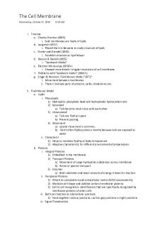
Exploration Research question What is the relationship between increasing ethanol concentration by increments of 20% and the diffusion of Betacyanin from beetroot cells into the external environment as the cell membrane is destroyed? Introduction Beetroot or Beta Vulgaris is a leafy plant with a dark red taproot. Its pigment is due to a naturally occurring red-violet compound called betanin. Betanin is a glycosidic molecule that is stored in the cell ’s large vacuole and is water-soluble. (Betanin, 2020) This vacuole is enclosed by a membrane called the tonoplast, which maintains the inner pressure in cells, so they don ’t become flaccid. Beetroot cells also have other important parts such as the cytoplasm, nucleus, cell wall and cell membrane. A cell membrane or plasma membrane is a semi-permeable barrier that surrounds the cytoplasm in living cells. This barrier separates the cell from its external environment. Cell membranes are especially important as they hold the cell’s major constituents such as the cytoplasm and organelles in place and prevent unwanted leakage. Besides this, they also play a major role in the regulation and transport of materials which enter the cell or leave the cell. As per the Fluid Mosaic model seen in Figure 1, membranes are comprised of a semi-fluid phospholipid bilayer, cholesterol only in animal cells and various proteins. ("Fluid-Mosaic Model", n.d.)
Figure 1: Fluid mosaic model describing the structure of a cell membrane ("Fluid-Mosaic Model", n.d.)
Phospholipids are lipids that are amphipathic, meaning they have both polar and non-polar parts. They have a polar head composed of glycerol and phosphate which is hydrophilic or capable of interacting with water and some other polar molecules. They also have two non-polar hydrophobic tails composed of which are incapable of dissolving or interacting with water, however, interact with other non-polar substances such as lipids. ("membrane | Definition, Structure, & Functions", n.d.)
Proteins are necessary as they allow the transport of molecules across the membrane, maintain structure, facilitate movement and act as receptors for cell signalling. Ethanol is a volatile, colourless and highly flammable organic chemical compound with the formula C2H5OH. It contains a methyl group, methylene group and a hydroxyl group. Ethanol is a polar molecule and is therefore hydrophilic, however it also has non-polar or hydrophobic properties due to its 2-carbon chain. (Coelho & Goldemberg, 2004) Due to this, ethanol is amphipathic and is capable of interacting with both water and lipids. Cell contact with ethanol results in distortion of the plasma membrane. This occurs due to ethanol being amphipathic, allowing it to penetrate through the membrane as it forms hydrogen bonds with phospholipids. (Patra et al., 2005) It also affects the fluidity of the membrane as it distorts proteins, resulting in increased permeability in the membrane. This destruction of the membrane along with the cell’s tonoplast results in Betanin leaking out of the cell. A colourimeter is a device which measures the absorbance of certain wavelengths of light in a solution. A solution will have pigment/colour if reflects a certain colour of visible light and absorbs all other light. In this case, betanin will reflect red light and absorb all other light, resulting in the deep red pigment visible to the eye. The colourimeter is capable of detecting and measuring this certain light reflection. (Roy Choudhury, 2014) Variables Dependant The dependant variable in this experiment was the diffusion of betanin from beetroot cells into their external environment after being damaged by ethanol. This was measured by determining the optical density of betanin-containing solutions using a colourimeter. Independent The independent variable in this experiment was the different ethanol concentrations ranging from 0%-100%, increasing in increments of 20%. These concentrations include 0% or distilled water, 20%, 40%, 60%, 80% and 100% or pure ethanol. Controlled 1. The size of each beetroot segment was controlled to be 0.5cm in width and 2cm in length so that each concentration and trial gave valid data for the same conditions. This was done through the use of cork borers to achieve the width and a ruler and knife to achieve suitable the length. 2. The type of alcohol used was a controlled variable because ethanol was used instead of methanol or 1-propanol. 3. The volume of ethanol and water solutions were controlled to ensure all trials underwent the same testing conditions. 5mL of each
concentration was measured using the graduated pipette and added to the test tube. 4. The submersion time for the beetroot in the solution was controlled to avoid variations in data caused due to unfair testing. This was carried out using a stopwatch to ensure each piece was in the solution for only 10 minutes. 5. The overall temperature and environment in which the experiment was conducted for each trial was controlled to increase the validity of data. This was done by conducting all trials in the same area and time period to avoid errors in the data. 6. The appropriate wavelength of 420nm was set on the colourimeter for the red pigment of betacyanin before use so that the data was valid and suitable.
Hypotheses H0- Ethanol has no effect on the structure of the beetroot membrane and no Betanin is released
H1- The relationship between ethanol concentrations and membrane destruction is directly proportional, meaning as the concentration of ethanol increases, more Betanin is released because it penetrates the beetroot membrane. Due to ethanol’s ability to denature cell membranes, it was hypothesised that an increase in ethanol concentration will result in more betanin pigment in the solution as the cell membrane is denatured. This hypothesis implies that 0% will have the least optical density, 20% will have more, 40% more and so on, with 100% having the most optical density indicating the most diffusion of Betanin in the cells’ surroundings. Equipment Colourimeter 1x ( 0.0001 mg/L) 3 mL disposable pipette 1x ( 0.05 mL) Weighing scale ( 0.001 g) Graduated pipette 1x ( 0.05 mL) Measuring cylinder 1x ( 0.05 mL) Stopwatch 1x Reagent bottles 6x Beaker of 100% ethanol 1x Test tube rack 2x Test tubes 18x Beetroot 1x Knife 1x Cutting board 1x Distilled water 1x Cuvette 6x Permanent marker 1x Cork borer 1x Ruler 1x
Methodology 1. The 100% ethanol was diluted into different concentrations including 20%, 40%, 60% and 80% using the graduated pipette. 2. Around 150mL of each concentration from 0-100% was made and placed in reagent bottles labelled with a permanent marker. 3. Using a graduated pipette 5mL of every ethanol concentration was extracted and placed in 3 separate test tubes on the racks for 3 different trials per concentration. 4. The beetroot was peeled and cut using the knife on the chopping board. Using a cork borer, a total of 18 cylinders of beetroot were bored. 5. Each piece was then measured and cut at 2cm using the knife. 6. All pieces were gently washed with distilled water to eliminate betacyanin that has already been leaked due to handling of beetroot. 7. A weighing scale was set up and 3 beetroot segments were chosen and weighed together for each concentration. This weight was recorded. 8. All 3 beetroot pieces were submerged into the separate 0% concentration test tubes at the same time and a timer was activated. The 0% concentration trials represent the negative control group. 9. At one minute intervals, the beetroot segments were consecutively submerged into the different ethanol concentrations. This was carried out until all concentrations contained a beetroot slice. 10. After 10 minutes, the beetroot pieces from 0% ethanol were removed together and the liquid from each test tube was mixed and moved to 3 corresponding cuvettes. 11. This was repeated consecutively after 1 minute intervals in order by which beetroot was first added to the ethanol concentrations. 12. All cuvettes were placed inside the colourimeter, which was plugged in and set to 420nm. The optical density for all trials associated with concentration was recorded to determine the amount of pigment leaked from beetroot for each concentration. 13. The beetroot pieces for each concentration were then reweighed together to obtain an after-experiment weight. Risk assessment Table 1: Table showing and explaining risks associated with conducting experiment and how they were controlled Risk Explanation How it was controlled Liquids were kept away from the Electrocuti There is a risk of devices and their plug-in cords on electrocution since were kept out of the way to electrical devices like a avoid tripping on them. colourimeter and weighing scale were used.
Glass injury
Glass equipment such as the graduated pipette and test tubes post a risk of cuts if broken.
Knife cuts
The use of a sharp knife to cut beetroot may have resulted in cuts to the person slicing. Ethanol is an irritant towards the skin as it dehydrates it. Contact with the eyes can result in burning and stinging.
Ethanol irritation
This was controlled by being careful when handling glass equipment. It was ensured they were placed correctly on the workspace or test tube racks to avoid breakage. The knife was handled carefully, and rubber gloves were worn to offer protection to hands. To avoid irritation, gloves were worn when handling ethanol and safety glasses were worn to avoid spraying of ethanol into the eye.
Analysis Qualitative data
0%
20%
40%
60%
80% 100%
Figure 2: Betanin and ethanol solutions for different concentrations increasing left to right
The observable changes that are shown in figure 2 display the pigment released by the beetroot for each concentration. It can be seen there is an increase in pigment intensity from 20% to 40% and from 60% to 80%. The pigment between 0% and 20% is not in favour of the hypothesis as the pigment decreases. The pigments 40% and 60% are also not in favour of the hypothesis as the intensity of the pigment stays about the same,
although there is a slight change in the colour. The 100% ethanol solution also had a visibly lower intensity of pigment compared to 80%.
Quantitative data Table 2: Quantitative processed data for the optical densities of betacyanin in different ethanol concentration solutions
Trial 3 ( Mean ( Std Trial 1 Trial 2 dev ( 0.001 0.003 ( ( 0.001 0.001 mg/L) 0.003 mg/L) mg/L) mg/L) mg/L) 0 0.109 0.065 0.106 0.0933 0.0245 20 0.105 0.071 0.062 0.0793 0.0226 40 0.180 0.310 0.267 0.252 0.0662 60 0.237 0.298 0.227 0.254 0.0384 80 0.518 0.412 0.291 0.407 0.113 100 0.466 0.258 0.255 0.326 0.121 Table 2 shows the measurement of the optical densities for each of the trials (1-3) of different ethanol concentrations that were measured using the colourimeter. The optical density indicates the amount of Betacyanin leaked from beetroot in each solution. The table indicates the data for the mean as well as individual trials of optical density for each concentration. It also with the standard deviation to show uncertainty.
Ethanol concentrati on (%)
Calculations for table 2 The following formula was used to calculate the mean optical densities of different ethanol concentrations. In this formula mean= m m=
∑ of terms number of terms
0% concentration: 0.109+0.065 + 0.106 m= 3 m=0.0933 mg/L 20% concentration: 0.105+0.071 + 0.062 m= 3 m=0.0793 mg/L 40% concentration: 0.180+0.310 + 0.267 m= 3 m=0.252 mg/L 60% concentration: 0.237+0.298 + 0.227 m= 3 m=0.254 mg/L
80% concentration: 0.518+0.412 + 0.291 m= 3 m=407 mg/L 100% concentration: 0.466+0.258 + 0.255 m= 3 m=0.326 mg/L The standard deviation for the different ethanol concentrations was calculated using an online calculator which has been referenced in the bibliography. ("Standard Deviation Calculator", n.d.) Figure 3: Mean optical density of leaked pigment from beetroot segments after 10 minutes of continuous contact with different ethanol concentrations ranging from 0%-100%
Mean optical density of leaked beetroot pigment for different ethanol concentrations 0.45 0.4
Optical density (mg/L)
0.35 0.3 0.25 0.2 0.15 0.1 0.05 0 0%
20%
40%
60%
80%
100%
Ethanol concentration (%)
It can be seen in figure 3 that the optical density increases only two sections like from 20% to 40% and from 60% to 80%. The density for 20% ethanol on the graph is lower than 0% according to the point, although there is a chance of this observation being unreliable due to the error bar. The optical density from 40% to 60% remains approximately constant and does not increase with concentration. There may also be possible changes in value due to the larger error bars seen from the 40% point. 80% ethanol has the highest optical density, suggesting that this concentration denatured the membrane the most as more betanin was released here than any other concentration. After 80%, the optical density once again decreases as it reaches 100% concentration. Although there is a high error rate for these two concentrations, the overall relationship between 80% ethanol and 100% ethanol upon membrane breakage seems valid. Table 3: Quantitative data for the mass of beetroot before and after continuous contact with different ethanol concentrations and change in mass
Ethanol
Mass of beetroot
Mass of
Change in
beetroot mass ( after ( 0.01 0.02 g) g) 0 1.13 1.40 0.27 20 1.23 1.26 0.03 40 1.20 1.18 0.02 60 1.09 1.02 0.07 80 1.13 0.92 0.21 100 1.12 0.61 0.51 Table 3 expresses the collective mass of all 3 beetroot pieces chosen for each concentration of ethanol both before and after performing the timed submersion. It then expresses the change in mass between the before and after masses. concentrati on (%)
before ( 0.01 g)
Calculations for table 3 The following formula was used to determine the change in mass for each ethanol concentration. Change in mass= c : c=mass after−mass before 0% concentration: c=1.40−1.13 c=0.27 g 20% concentration: c=1.26−1.23 c=0.03 g 40% concentration: c=1.18−1.20 c=−0.02 g 60% concentration: c=1.02−1.09 c=−0.07 g 80% concentration: c=0.92−1.13 c=−0.21 g 100% concentration: c=0.61−1.12 c=−0.51 g Figure 4: Change in mass of beetroot segments before and after 10 minutes of continuous contact with ethanol concentrations ranging from 0%-100%
Change in mass of beetroot segments for different ethanol concentrations 0.4 0.27 0.3
Change in mass (g)
0.2 0.1
0.03
0 0% -0.1
20%
-0.02 40%
-0.07 60%
80%
100%
-0.21
-0.2 -0.3 -0.4
-0.51
-0.5 -0.6
Ethanol concentration (%)
Figure 4 shows the change in mass between the before and after masses for each ethanol concentration. The mass for 0% and 20% after contact with ethanol solution was increased since figure 4 shows these values as a positive change in mass. However, the graph shows all concentrations for 40% or higher to have a negative change in mass. This indicates that the beetroot pieces for these concentrations lost mass. It can be seen that mass decreases more as the concentration increases since there is a change of 0.02g for 40%, 0.07g for 60%, 0.21g for 80% and 0.51g for 100%.
Evaluation The purpose of this report was to contribute knowledge on the effect of ethanol upon cell membranes. The experiment carried this by testing on beetroot slices with varying concentrations of ethanol. The experiment’s helps to answer the research question and to determine whether or not the hypotheses were supported. There is a link between The data concluded that increasing the ethanol concentration in increments of 20% does not result in more betacyanin diffusion due to damage to the membrane. It can be seen that the data does not support the hypothesis. According to hypothesis1 the ethanol concentrations and optical density should have a directly proportional relationship, meaning as concentration increases the amount of pigment in the solution should increase accordingly. Looking at Figure 3, some observations according to the hypothesis were seen between 20% (0.0793 mg/L) to 40% (0.252 mg/L) concentrations and 60% (0.254 mg/L) to 80% (0.407 mg/L) concentrations since these are the only areas where pigment leakage increases along with the concentration. However, for the most part it can be seen that there is not a directly proportional relationship between the
ethanol concentration and betacyanin diffusion as between 0% to 20%, 40% to 60% and 80% to 100% there is almost no proportional increase. Instead, there is a very minimal decrease in optical density between 0% to 20% by 0.02 mg/L. Alongside, there is a significant decrease of optical densities between 80% to 100% by 0.81 mg/L. Due to these observations, hypothesis1 was not supported.
Restate findings and explain all turning points on graphs DISCUSSIONNegative control or 0% concentration Negative control- everything but dependant (0% or just water) Limitations: Same pipette used for every concentration and was not calibrated Beetroot pieces were not all taken out at exact same times Precision- std dev Validity- negative control Accuracy- theory
Bibliography Betanin. (2020). Retrieved 4 September 2020, from https://en.wikipedia.org/wiki/Betanin Patra, M., Salonen, E., Terama, E., Vattulainen, I., Faller, R., & Lee, B. et al. (2005). Under the Influence of Alcohol: The Effect of Ethanol and Methanol on Lipid Bilayers. Retrieved 8 September 2020, from https://www.ncbi.nlm.nih.gov/pmc/articles/PMC1367264/ Standard Deviation Calculator. Retrieved 8 September 2020, from https://www.calculator.net/standard-deviation-calculator.html Fluid-Mosaic Model. Retrieved 9 September 2020, from https://ib.bioninja.com.au/standardlevel/topic-1-cell-biology/13-membrane-structure/fluid-mosaic-model.html Coelho, S., & Goldemberg, J. (2004). Ethanol - an overview. Retrieved 9 September 2020, from https://www.sciencedirect.com/topics/earth-and-planetary-sciences/ethanol Roy Choudhury, A. (2014). Colorimeter - an overview. Retrieved 10 September 2020, from https://www.sciencedirect.com/topics/engineering/colorimeter...
Similar Free PDFs
Popular Institutions
- Tinajero National High School - Annex
- Politeknik Caltex Riau
- Yokohama City University
- SGT University
- University of Al-Qadisiyah
- Divine Word College of Vigan
- Techniek College Rotterdam
- Universidade de Santiago
- Universiti Teknologi MARA Cawangan Johor Kampus Pasir Gudang
- Poltekkes Kemenkes Yogyakarta
- Baguio City National High School
- Colegio san marcos
- preparatoria uno
- Centro de Bachillerato Tecnológico Industrial y de Servicios No. 107
- Dalian Maritime University
- Quang Trung Secondary School
- Colegio Tecnológico en Informática
- Corporación Regional de Educación Superior
- Grupo CEDVA
- Dar Al Uloom University
- Centro de Estudios Preuniversitarios de la Universidad Nacional de Ingeniería
- 上智大学
- Aakash International School, Nuna Majara
- San Felipe Neri Catholic School
- Kang Chiao International School - New Taipei City
- Misamis Occidental National High School
- Institución Educativa Escuela Normal Juan Ladrilleros
- Kolehiyo ng Pantukan
- Batanes State College
- Instituto Continental
- Sekolah Menengah Kejuruan Kesehatan Kaltara (Tarakan)
- Colegio de La Inmaculada Concepcion - Cebu


