Notes Chapter 13 Spinal Cord and Spinal Nerves PDF

| Title | Notes Chapter 13 Spinal Cord and Spinal Nerves |
|---|---|
| Author | jared smith |
| Course | Human Anatomy and Physiology |
| Institution | Athabasca University |
| Pages | 11 |
| File Size | 579.1 KB |
| File Type | |
| Total Downloads | 77 |
| Total Views | 208 |
Summary
Download Notes Chapter 13 Spinal Cord and Spinal Nerves PDF
Description
Biology 235 – Athabasca University
October 1 2020
Chapter 13: Spinal Cord and Spinal Nerves
Spinal Cord Anatomy -skull and vertebral column act as 1st layer of protection for CNS -2nd layer of protection are the meninges, three membranes that lie between the bony skull and vertebrae and the nervous tissue of the brain and spinal cord -3rd layer protection is the cerebrospinal fluid (CSF), in between the inner two meninges, which suspends nervous tissue in weightless environment and provides shock absorbing cushion
3 Meninges (singular: meninx) 1. Dura Mater (most superficial / outer): thick, strong layer of dense irregular connective tissue 2. Arachnoid (middle layer): thin, avascular covering of loosely arranged collagen and elastic fibres. In between dura mater and arachnoid is subdural space which contains interstitial fluid 3. Pia Mater (innermost layer): thin, transparent connective tissue layer that adheres to surface of spinal cord and brain. Made up of vascular squamous to cuboidal cells with collagen and elastic fibres. Has blood vessels which supply oxygen and nutrients to spinal cord. In between arachnoid layer and pia mater is the subarachnoid space which contains cerebrospinal fluid
External Anatomy of Spinal Cord -Spinal cord extends from medulla oblongata (inferior part of brain) to the superior border of the 2nd lumbar vertebra (in adults) Cervical enlargement: enlargement of the spinal cord between C4 and T1. Nerves to and from the upper limbs arise Lumbar enlargement: from 9th to 12th thoracic vertebra. Nerve to and from lower limbs arise from here. Conus medullaris: the tapered, conical ending of the spinal cord, inferior to the lumbar enlargement. Ends at intervertebral disc between 1st and 2nd lumbar vertebrae Filum terminale: extension of the pia mater that goes from conus medullaris and anchors spinal cord to coccyx Spinal nerves: pathways of communication between spinal cord and specific regions of body
31 pairs of spinal nerves: 8 cervical, 12 pairs thoracic, 5 pairs lumbar, 5 pairs sacral, and 1 pair coccygeal nerves Cauda equine: nerves that do not leave the vertebral canal in the same place they leave the spinal cord, instead they travel inferiorly towards the filum terminale
Internal Anatomy of the Spinal Cord
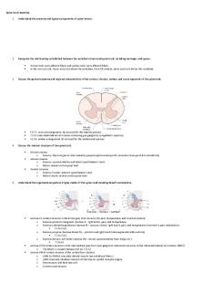
white (myelinated) matter surrounds inner core (H shaped) of gray matter (unmyelinated) o white matter is mostly bundles (tracts) of myelinated axons of neurons o gray matter mostly dendrites and cell bodies of neurons, unmyelinated axons, and neuroglia
in gray matter, clusters of neuronal cell bodies form nuclei. Sensory nuclei receive input from receptors via sensory neurons and motor nuclei provide output to effector tissues via motor neurons
1. anterior median fissure: groove on anterior side
posterior median sulcus: narrow furrow on posterior side grey commissure: cross-bar of H central canal: extends entire length of spinal cord. Filled with cerebrospinal fluid anterior white commissure: nuclei: clusters of neuronal cell bodies in gray matter posterior gray horn: contains cell bodies and axons of interneurons as well as axons of incoming sensory neurons 8. anterior gray horn: contain somatic motor nuclei, cluster of cell bodies of somatic motor neurons tat provide nerve impulses for contractions of skeletal muscle 9. lateral grey horn: contain autonomic motor nuclei that regulate activity of cardiac and smooth muscle and glands. 10. Columns: white matter divided into columns. Each contain bundles of axons that have common origin or destination and carrying similar information 11. anterior white column 12. posterior white column 13. tract: bundle of axons in CNS 14. sensory (ascending) tract: axons conducting nerve impulses towards brain 15. Motor (descending) tract: axons conducting nerve impulses from the brain to effector muscles or glands 2. 3. 4. 5. 6. 7.
How Spinal Nerves are connected to Spinal Cord (see diagram) Two bundles of axons, called roots, connect each spinal nerve to spinal cord by even smaller bundles called rootlets Each of the spinal nerves divides into two roots and enters the spinal cord o Posterior root, anterior root, and posterior root ganglion (enlargement of nerve right after nerve divides into two roots.
Spinal Nerves (part of the PNS) spinal nerves are parallel bundles of axons (and neuroglia) within layers of connective tissue Spinal nerves connect CBS to sensory receptors, muscles, and glands in all parts of the body 31 pairs of spinal nerves named and numbered by which region of the vertebral column they emerge from Typical spinal nerve has two connections to spinal cord: posterior root and anterior root Because posterior root contains sensory axons and anterior root contains motor axons, spinal nerves are classified as mixed nerves
Connective Tissues Covering Spinal Nerves 4 layers: (inner to outer) endoneurium, fasciles, perineurium, epineurium
each spinal nerve made up of many individual axons individual axons (myelinated or unmyelinated) are wrapped in endoneurium (innermost layer) made of collagen fibres, fibroblasts, and macrophages groups of axons held together in bundles called fascicles, each of which is wrapped in perineurium (middle layer) which is also the thickest layer (up to 15 layers of fibroblasts) Outermost covering is epineurium which is vascular
Branches of Spinal Nerves
After passing through intervertebral foramen, spinal nerves divide into several branches called rami (ramus = singular). Spinal nerve divides into (3): o Posterior ramus serves deep muscles and skin of the posterior surface of the truck o Anterior ramus serves muscles and structures of upper and lower limbs and the skin of the lateral and anterior surfaces of the trunk o Meningeal branch: which re-enters the vertebral cavity through intervertebral foramen and supplies vertebrae, vertebral ligaments, blood vessels of spinal cord, and meninges
Plexuses (see big diagram above)
Axons from anterior rami of spinal nerves to not go directly to body part. They first go to networks of axons from adjacent nerves which are called a plexus There are 5 main plexuses: cervical, brachial, lumbar, sacral, coccygeal From a plexus emerge nerves with names that often describe the regions they serve or course they take
Intercostal Nerves T2 – T12 do not go into plexuses. Instead, they connect directly to the structures they supply in the intercostal spaces.
T2 innervates intercostal muscles of the 2nd intercostal space. Also skin of axilla and posteromedial aspects of arm T3 – T6 innervate intercostal muscles and skin of anterior and lateral chest wall T7 – T12 innervates intercostal muscles and abdominal muscles along with skin Posterior rami of intercostal nerves innervate deep back muscles and skin of posterior thorax
Dermatome: an area of skin that provides sensory input to the CNS via one pair of spinal nerves of the trigeminal nerve (face and scalp). *See figure 12.11 which is a map of the dermatomes
Nerve supply in adjacent dermatomes overlap a little bit Knowing which spinal nerve innervates each dermatome makes it possible to locate damaged regions of the spinal cord.
Functions of Major Sensory and Motor Tracts of the Spinal Cord
Spinal cord has two major functions in maintaining homeostasis o Nerve impulse propagation o Integration (processing) of information
The white matter tracts in spinal cord are highways for nerve impulse propagation o Sensory inputs travel from sensory neuron towards brain and motor output from brain travels along them towards skeletal muscle and other effector tissues (glands)
Gray matter of spinal cord receives and integrates incoming and outgoing information
Sensory and Motor Tracts
Name of tract sometimes indicates its position in the white matter and where it beings and ends in body. o Example: anterior corticospinal tract is located in the anterior of the white column; it begins in the cerebral cortex and ends in the spinal cord (location of axon terminals come last in name) o Because it conveys info away from the brain, it is a motor (descending) tract
Sensory Tracts (2 main) (ascending)
Spinothalamic tract: conveys nerve impulses for sensing pain, warmth, coolness, itching, tickling, deep pressure, and crude touch up spinal cord to brain Posterior Column: conveys nerve impulses for discriminative touch, light pressure vibrations, and conscious proprioception (awareness of position and movement of muscles, tendons, and joints). Consists of two tracts: o Gracile fasciculus
o Cuneate fasciculus -sensory systems tell CNS of changes in internal and external environment. Sensory information integrated (processed) by interneurons in spinal cord and brain. Responses to that processing are made by motor activities (muscular contractions and glandular secretions).
Motor Tracts (descending)
Direct motor pathways: convey nerve impulses that will create voluntary movements of skeletal muscles Indirect motor pathways: convey nerve impulses that will cause automatic movements and help coordinate body movement with visual stimuli. o also maintain body tone, contraction of postural muscles, and regulate muscle tone in movements of head
Reflex and Reflex Arc -another way (in addition to conducting nerve impulses) that spinal cord helps maintain homeostasis is by acting as an integration (processing) centre for some reflexes. Reflex: a fast, involuntary, unplanned sequence of actions that occur in response to a stimulus. Some innate (inborn): pulling hand away from hot surface; other are learned: hitting car brakes in emergency When integration occurs in spinal cord, it is a spinal reflex (i.e. patellar reflex) If integration occurs in brain, then it’s a cranial reflex Somatic reflexes involve contraction of skeletal muscles Autonomic (visceral) reflexes: usually not conscious (i.e. smooth muscle, cardiac muscle, glands)
Reflex arc (reflex circuit): (see figure 13.13)
the pathway followed by a nerve impulse that produces a reflex Reflex arc includes following 5 components 1. Sensory Receptor: distal end of a sensory neuron (dendrite) or other sensory structure. Responds to stimulus by producing graded potential called generator potential (or receptor potential). If it reaches threshold, it will initiate a nerve impulse in the sensory neuron 2. Sensory Neuron: nerve impulse propagates from sensory receptor along axon of sensory neuron to the axon terminals which are in the gray matter of the spinal cord or brain. 3. Integrating Centre: one or more regions of CNS act as integrating centre.
o Monosynaptic reflex (most simple): integrating centre is a single synapse between a sensory neuron and a motor neuron o Polysynaptic reflex: integrating centre is more than one neuron that may relay impulses to other interneurons as well as to motor functions. 4. Motor neuron: impulse triggered by integrating centre propagate out of CNS along motor neuron to the part of the body that will respond (the effector) 5. Effector: the part of the body that responds to the motor nerve impulse (muscle or gland). Its action is called a somatic reflex o If effector is smooth muscle, cardiac muscle, or a gland, the reflex is an autonomic (visceral) reflex
Stretch reflex: causes contraction of a skeletal muscle in response to stretching a muscle. Occurs via monosynaptic reflex arc. Stretch reflex can be elicited by tapping on tendons on attached to elbow muscles, wrist, knee, and ankle joints. One example is patellar reflex (knee jerk) *see figure 13.14 How it works: 1. Stretching of muscle stimulates sensory receptors called muscle spindles that sense change in length of muscle 2. In response to stretch, muscle spindle generates nerve impulse along somatic sensory neuron, through posterior root of spinal nerve, and into the spinal cord. 3. In spinal cord (integrating sensor), sensory neuron makes excitatory synapse with a motor neuron in the anterior gray horn 4. If it reaches threshold, nerve impulse propagates along motor neuron, through anterior root and peripheral nerve to stimulated muscle 5. Acetylcholine released by nerve impulse at the NMJ triggers muscle action potentials and the stretched muscles contracts. Muscle stretch is followed by muscle contraction which relieves the stretching
Ipsilateral reflex arc: when sensory nerve impulses enter spinal cord on the same side from which motor nerve impulses leave it. All monosynaptic reflexes are ipsilateral
By adjusting how much a muscle spindle responds to stretching, brain sets an overall level of muscle tone (the amount of contract present when muscle at rest) Stretch reflex is monosynaptic (just two neurons and one synapse), but a polysynaptic reflex arc to antagonist muscle operates at the same time. This inhibits a nerve impulse from travelling to the antagonistic muscle. Stretch reflex helps maintain posture. For example, standing person leaning forward, calf muscles are stretched. Stretch reflex causes them to contract which brings the person back into balance This is called reciprocal innervation, when components of a neural circuit cause contraction of one muscle and relaxation of its antagonist. Prevents conflict between opposing muscles
Tendon reflex: causes muscles to relax before muscle force becomes great enough that something tears (like a tendon). It’s a feedback mechanism that controls muscle tension. Receptors for this reflex called tendon organs (golgi tendons) (within tendon near junction with muscle). Monitors changes in muscle tension due to passive stretch or muscular contraction. How it works: 1. As tension applied to tendon increases, tendon organ (receptor) is stimulated (depolarized to threshold) 2. Nerve impulse propagated into spinal cord along sensory neuron 3. Spinal cord (integrating centre), sensory neuron activates inhibitory interneuron that synapses with motor neuron
4. Inhibitory neurotransmitter inhibits (hyperpolarizes) the motor neuron, which then generates fewer nerve impulses 5. The muscle relaxes and relieves excess tension
Flexor reflex (withdrawl reflex): (example: withdraw leg after stepping on tack) 1. 2. 3. 4. 5.
Stepping on tack stimulates dendrites (sensory receptor) of pain sensitive neuron This sensory neuron generates nerve impulses which travel to spinal cord Sensory neuron activates interneurons in spinal cord Interneurons activate motor neurons in several spinal cord areas and initiate nerve impulses Acetylcholine released by motor neurons causes flexor muscle in thigh to contract, withdrawing leg It is a polysynaptic reflex arc b/c many muscles are needed to pull leg away. It’s also an intersegmental reflex because a single sensory neuron ascend and descend spinal cord activating interneurons in several areas.
Crossed extensor reflex: helps you maintain balance. Sends nerve impulses to other leg to contract thereby allowing you to put all your weight on that leg as you unweight the leg which you stepped on the tack with Contralateral reflex arc: sensory impulses enter one side of the spinal cord and motor impulses exit on the opposite side (as in crossed extensor reflex)
Patellar reflex: stretch reflex that cuases extension of the leg at knee (contraction of quadricep femoris) in response to tapping on patellar ligament Babinski reflex: reflex results from stroking of the lateral outer margin of the sole. Causes great toe to extend. Normal in children under 1.5 years old, after that, it indicates interruption of corticospinal tract. A negative Babinski test (indicating everything is normal) would see the patient’s toes curl under.
Disorders:
1. Spinal cord compression: can result from fractured vertebrae, herniated intervertebral discs, tumours, osteoporosis, or infections. Symptoms include pain, weakness or paralysis, and loss of sensation below level of the injury. 2. Shingles: acute infection of PNS cause by herpes zoster virus (same that causes chickenpox). Symptoms: pain, discoloration of skin, and line of skin blisters. 3. Poliomyelitis (polio): caused by virus called poliovirus. Onset is marked by fever, headache, stiff neck and back, loss of certain somatic reflexes, and deep muscle pain and weakness. Can cause paralysis if it destroys cell bodies of motor neurons....
Similar Free PDFs

Ch17 Spinal Cord Nerves
- 4 Pages

ANS and Spinal Cord Labeling
- 3 Pages

Spinal cord, medulla spinalis
- 5 Pages

Spinal Cord and Lower Brain
- 5 Pages

Spinal Cord Injury
- 1 Pages

Spinal Nerves SI - Hargroder
- 4 Pages
Popular Institutions
- Tinajero National High School - Annex
- Politeknik Caltex Riau
- Yokohama City University
- SGT University
- University of Al-Qadisiyah
- Divine Word College of Vigan
- Techniek College Rotterdam
- Universidade de Santiago
- Universiti Teknologi MARA Cawangan Johor Kampus Pasir Gudang
- Poltekkes Kemenkes Yogyakarta
- Baguio City National High School
- Colegio san marcos
- preparatoria uno
- Centro de Bachillerato Tecnológico Industrial y de Servicios No. 107
- Dalian Maritime University
- Quang Trung Secondary School
- Colegio Tecnológico en Informática
- Corporación Regional de Educación Superior
- Grupo CEDVA
- Dar Al Uloom University
- Centro de Estudios Preuniversitarios de la Universidad Nacional de Ingeniería
- 上智大学
- Aakash International School, Nuna Majara
- San Felipe Neri Catholic School
- Kang Chiao International School - New Taipei City
- Misamis Occidental National High School
- Institución Educativa Escuela Normal Juan Ladrilleros
- Kolehiyo ng Pantukan
- Batanes State College
- Instituto Continental
- Sekolah Menengah Kejuruan Kesehatan Kaltara (Tarakan)
- Colegio de La Inmaculada Concepcion - Cebu


