Spinal Cord and Lower Brain PDF

| Title | Spinal Cord and Lower Brain |
|---|---|
| Author | Kamryn Stone |
| Course | Human Anat & Physiol I |
| Institution | University of Louisville |
| Pages | 5 |
| File Size | 91.3 KB |
| File Type | |
| Total Downloads | 73 |
| Total Views | 203 |
Summary
This lecture was led by Jennifer Mansfield-Jones and cover the topics of the Spinal Cord and Lower Brain...
Description
Spinal Cord and Lower Brain The CNS Development ● The CNS begins as a hollow tube on the dorsal side of the embryo ○ Explains a lot about the setup of the whole CNS of the adult ● At just short of 3 weeks we have a development of a flat spot that develops ridges that rise up ○ Neural folds that come up and come together to form the hollow “tube” ■ Made up of the tissue that used to be the “skin” of the embryo ● Neural Tube: Where the CNS is formed in an embryo ○ Can have defects - inadequate intake of folic acid ● Neural Crest Cells: The crest of the ridges that form the neural tube ○ They do a lot of jobs - can become melanocytes in the skin & forms ganglia in the PNS ● Fetal Alcohol Syndrome - Ethanol interfering with the normal travels of the NCCs ○ Heart problems and irregular facial features
Tube-Like Spinal Cord ● The walls of the hollow tube thickens and contains a lot of neurons and glial cells - still a hollow tube though ○ The “hole” in the spinal cord is called the central canal ● Central Canal: The hole that the spinal cord is fed through ● The spinal cord is not the same thickness ○ 1) Thicker in the lower cervical region - Cervical enlargement ○ 2) Enlarges again in the thoracic region - Lumbar enlargement (but not located in the lumbar area) ○ The end of the spinal cord looks like a sharp point and is very narrow ● Spinal cord is not the entire length of the vertebral column ● Cauda Equina: (horses tail) Nerve roots at the end of the spinal cord that arrange in coarse strings ○ NOT PART OF CENTRAL NERVOUS SYSTEM ○ Part of PNS ● The body of the vertebrae protects the front of the spinal cord & the spinous process and pedicle protect the back ○ Pedicles also protect the sides ● Spinal nerve roots are fed through the foramina of the vertebrae ● Additional Protection: A set of three layers - Meninges ○ 1) Pia Mater - Delicate and innermost layer ○ 2) Arachnoid - Middle layer ○ 3) Dura Mater- Outermost and toughest layer
● Arachnoid layer has a layer of fluid found within it (cerebro-spinal fluid) ○ Fluid padding for the spinal cord ○ Dura Mater keeps everything on the inside all “snuggly” in place ● Between the meninges and the bone there is more padding - Epidural Space ○ Full of fat (adipose tissue padding) ● Ex: Packaging a fragile item in a sturdy box ○ Item in the middle encased by several “fluff” structures to keep it in place and from getting damaged
Meninges Around Spinal Cord ● Organization from innermost to outermost: ○ Pia Mater - Thin and innermost layer ○ Arachnoid - Middle layer that contains fluids for padding ○ Dura Mater - The toughest and outermost layer that holds everything together ● Roots of nerves connect directly to the spinal cord via rootlets ○ Rootlets: The ending of the nerve that connects to the spinal cord ● Spinal cord has grooves on anterior and posterior sides ○ Anterior: Deeper and more obvious - Anterior Median Fissure ■ Is generally flat ○ Posterior: A less dramatic groove in the spinal cord -Posterior Median Sulcus ■ Bulgey instead of flat ● Spinal nerve roots are also good indicators of which way the spinal cord is being shown: ○ Anterior: Ventral roots are smooth ○ Posterior: Dorsal roots have a large bulge in them (dorsal root ganglion)
Spinal Cord Anchoring ● Central Canal: A hole full of cerebrospinal fluid ○ Two places with this fluid, 1) The Central Canal 2) The Arachnoid Layer ● Anchoring Mechanisms: ○ 1) The layers of Meninges - Pia Mater, Arachnoid, and Dura Mater ○ 2) Denticulate Ligaments - Connective tissue that further anchors the spinal cord from the sides ■ Run from the Pia Mater out through the Arachnoid Layer and out to the Dura Mater ■ The name “Denticulate” refers to a tooth ○ 3) Epidural Adipose Tissue - A layer of fat that helps for padding
Different Neurons in Different Places
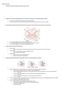
● Ganglion = Group of neuron cell bodies in PNS ● Nucleus = Group of neuron cell bodies in CNS ● Dorsal Root Ganglion - Where the sensory neuron bodies are locates ○ DRG connect to Dorsal Root fibers that connect to the posterior side of the spinal cord ● “Paler” nervous tissue shown in spine x-ray is called White Matter (yellow/white because of presence of myelin) ○ Oligodendrocytes ● “Butterfly” shaped tissue in the middle of the spinal cord is called Grey Matter (Unmyelinated) ○ Neuron cell bodies found here ○ Posterior side of butterfly - Dorsal Grey Horns ○ Anterior side of butterfly - Ventral/Anterior Grey Horns ■ We find neuron cell bodies that belong to Somatic Motor Neurons here ● The axons of these SMNs go out through the ventral roots of the spinal nerves
Spinal Cord White Matter ● Motor pathways are not limited to one side of the spinal cord, but are often depicted this way to avoid confusion ● Nerve = Group of neuron axons running together in PNS ● Root/Rootlet = Smaller group joining others to form nerve ● Tract = Group of neuron axons running together in CNS ● All of the posterior white matter of the spinal cord consists of sensory tracts ○ Carrying sensory information fast up the spinal cord to the brian ● Lateral/Anterior - Ascending (sensory) and descending (motor) pathways
Sensory and Motor Pathways ● Incoming Sensory Information Pathway: ○ Sensory Neuron (unipolar shape) recognizes a stimulus and fires an action potential - Bringing information in ○ Interneuron (second order neuron) accepts the action potential from the sensory neuron ■ Axon crosses over to the other side of the spinal cord (shifting the information to the opposite side of CNS) - decussation ○ Axon goes up on the opposite side of the SC into the brain (opposite side of the stimulus) ■ Goes to the thalamus - The center that decides whether or not we are conscious of incoming information ○ From the thalamus the information is transferred to another interneuron (third-
order neuron) ● Motor Pathway: ○ Upper part of the brain considers the stimulus and has come to the conclusion some motion needs to take place ■ Triggers the activation of interneuron in the conscious part of our brains that send information down to the deeper parts of our brains and then into the spinal cord ○ The crossing over of the information also happens here (decussation) ○ The information is then carried out and the motion/movement happens
Brain Formation ● Starts out as a neural tube just like the spinal cord, but gets a more complex shape ● As the brain develops the front part of the NT get larger and the very front part develops a fork or two arms (telencephalon) ○ Telencephalon - The part of the brain that forms the highest conscious part of our brains, the cerebral hemispheres ● Diencephalon - Where the structures that will form the retinas of our eyes grow out from (little forked structures) ● Mesencephalon = Midbrain - A landmark structure that differentiates forebrain structures from hindbrain structures ● Metencephalon - In between the Mesencephalon and the Medulla Oblongata ● Medulla Oblongata - Region of the brain important for a lot of vegetative sort processes, and the passage of information from the brain to the spinal cord ● As the telencephalon part of the brain expands and folds down over the rest, it conceals a lot of the more inferior parts of the brain
Brain Ventricles ●
Visible from the outside: ○ Spinal Cord ○ Medulla Oblongata ○ Pons ○ Cerebellum ○ Cerebral Hemispheres ● Fluid filled spaces inside of the brain ○ If enlarged they’re called Ventricles ● At the bottom of the medulla oblongata and the cerebellum - Fourth Ventricle ○ Not really large and it tapers down to a narrow structure (cerebral aqueduct) ○ Cerebral Aqueduct passes right through the midbrain ● Third Ventricle looks large from a lateral view but not from an anterior view (skinny and flat)
● ●
● ● ● ●
○ Has structures of the thalamus on each side of it - useful for finding thalamus The Lateral Ventricles are located inside the cerebral hemispheres ○ They are curved because the structure they reside in are also curled The Medulla Oblongata connects to the spinal cord ○ Top of Medulla Oblongata is indicated by a bulge on the front side ■ Anterior - Pons starts ■ Posterior - Where the cerebellum is Cerebellum is finely folded and is unique - A really good landmark of the brain Mid-Brain sits above the Pons and cerebellum Diencephalon sits above the midbrain Telencephalon wraps around the whole brian structure
Brain Protection ● The brian contains the same Meninges as the spinal cord ○ Pia Mater, Arachnoid, and Derma Mater ● The brain makes these cerebrospinal fluids ○ Choroid Plexus Blood Vessels - constantly make new cerebrospinal fluid ■ Between ependymal cells and blood vessels we filter out new cerebrospinal fluid ○ This must be filtered out to avoid making too much fluid ■ Recycling happens through valve-like structures - Arachnoid Villi ○ If not recycled pressure will build up and have an enlarged cranium...
Similar Free PDFs

Spinal Cord and Lower Brain
- 5 Pages

ANS and Spinal Cord Labeling
- 3 Pages

Spinal cord, medulla spinalis
- 5 Pages

Spinal Cord Injury
- 1 Pages

Ch17 Spinal Cord Nerves
- 4 Pages
Popular Institutions
- Tinajero National High School - Annex
- Politeknik Caltex Riau
- Yokohama City University
- SGT University
- University of Al-Qadisiyah
- Divine Word College of Vigan
- Techniek College Rotterdam
- Universidade de Santiago
- Universiti Teknologi MARA Cawangan Johor Kampus Pasir Gudang
- Poltekkes Kemenkes Yogyakarta
- Baguio City National High School
- Colegio san marcos
- preparatoria uno
- Centro de Bachillerato Tecnológico Industrial y de Servicios No. 107
- Dalian Maritime University
- Quang Trung Secondary School
- Colegio Tecnológico en Informática
- Corporación Regional de Educación Superior
- Grupo CEDVA
- Dar Al Uloom University
- Centro de Estudios Preuniversitarios de la Universidad Nacional de Ingeniería
- 上智大学
- Aakash International School, Nuna Majara
- San Felipe Neri Catholic School
- Kang Chiao International School - New Taipei City
- Misamis Occidental National High School
- Institución Educativa Escuela Normal Juan Ladrilleros
- Kolehiyo ng Pantukan
- Batanes State College
- Instituto Continental
- Sekolah Menengah Kejuruan Kesehatan Kaltara (Tarakan)
- Colegio de La Inmaculada Concepcion - Cebu


