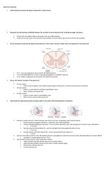
*Spinal cord and nerves - Google Docs PDF

| Title | *Spinal cord and nerves - Google Docs |
|---|---|
| Author | Aloe Bera |
| Course | Human Anatomy and Physiology I |
| Institution | McMaster University |
| Pages | 10 |
| File Size | 273.7 KB |
| File Type | |
| Total Downloads | 35 |
| Total Views | 197 |
Summary
Download *Spinal cord and nerves - Google Docs PDF
Description
Spinal cord ● Long structure comprised of nervous tissue (neurons and associated glial cells) ● Part of the CNS ● Located in the vertebral canal ● Functions of the spinal cord: Vertebrae- bone structures that surround the spinal cord ● Stacked on each other to create the vertebral column. ● Smooth side of the vertebrae = anterior side ● Spinous processes side =posterior side ● Function: provide protection for the tissues of the spinal cord Vertebral foramen- a hole within the spinous processes region that faces superior inferior ● When vertebrae are stacked on top of each other, the successive holes create a canal known as the vertebral canal. ● The vertebral column is divided into regions based on it's curves (superior -> inferior) ○ Cervical region ■ Cervical vertebrae ■ Cervical nerves ○ Thoracic region ○ Lumbar region ○ Sacral and Coccygeal regions ● The name of the regions dictate the names of the vertebrae and the spinal nerves (the spinal nerves that exit) in that region. Intervertebral foramina- spaces between the vertebrae ● Where the spinal nerves exit the vertebral canal.
● Spinal cord starts at the foramen magnum (Circular shaped hole in the base of the skull) and ends by the time it reaches the top of vertebrae L2 (second lumbar vertebrae) ○ Vertebral canal is longer than the spinal cord due to difference of speed of growth during development ● The spinal cord is kept in place in the middle of the spinal column by the meninges (connective tissue layers) ● Spinal cord in not quite round, it is flattened on one side ● Diameter of the spinal cord is not uniform along it's length. ○ There are two enlargements : 1. Cervical enlargement ● Occurs between vertebra C4 & T1 2. Lumbosacral enlargement ● Occurs between vertebrae T9 & T12 ○ The enlargements are for extra nervous tissues that are required to supply additional structures of the upper and lower limbs, as they require more neurons Conus medullaris- The inferior end of the spinal cord. ● Spinal cord tapers into a cone shape ● Nerves supplying the lower limbs exit the lumbosacral enlargement and course down through the vertebral canal to exit via the foramina. Coda equina- the hair like nature of the nerves coming out of the bottom of the spinal cord via the foramina
Filum Terminale- extension of the Pia mater connective tissue around the spinal cord. Anchors the spinal cord in the inferior part of the vertebral column. Stops any movement of the spinal cord in the superior direction
Meninges ● The spinal cord is surrounded by meninges to provide some level of protection. ● Continuous of the meninges of the brain. ● Layers of the meninges (superficial – deep): ○ Dura Mater ● Made up of irregular connective tissue ● Thickest and strongest layer ● There is space, Epidural space, between the dura mater and the periosteum (connective tissue on the bone) ● Space is filled with fat, blood vessels, areolar connective tissue ● Continuus with the brain ● Prevents the spinal canal from moving ○ Arachnoid Mater ● Thin, avascular (no blood supply to the layer) layer ● Contains simple squamous epithelial cells, and delicate network of collagen and elastic fibres. Subdural space: space between the dura and arachnoid mater. Contains small amount of serous fluid. ○ Pia Mater ● Contains blood vessels that supply the spinal cord ● Tightly adheres to the spinal cord ● Maintains the spinal cord in the vertebral canal ● Has small extensions of itself called the denticulate ligaments, that connect it to the dura mater.
● Denticulate ligaments also help anchor the spinal cord laterally to prevent side to side movement. Subarachnoid space- space between the pia mater and the arachnoid mater. Contains CSF. Continuus with the subarachnoid space of the brain ● The cerebral spinal fluid (CSF) helps cushion the spinal cord, delivers nutrients and removes waste products.
Internal anatomy ○ The spinal cord has two major portions: ○ Outer white matter portion ○ Inner grey matter portion
Posterior median sulcus- small groove on the posterior side of the spinal cord Anterior median fissure- large groove on the anterior side of the spinal cord ● The sulcus and the fissure divide the spinal cord into two halves.
White matter ● Contain myelinated axons which run the length of the spinal cord ● Can be ascending or descending creating nerve tract. ● Nerve tracts with similar functions are grouped in an organized way and become known as columns. ● These allow for information to be transmitted in an organized fashion throughout the spinal cord. ● There are 3 columns in the white matter: ○ Dorsal (Posterior) column ○ Ventral (Anterior) column ○ Lateral column White commissure- Connects the right and left side of the spinal cord. There are two of them, an anterior and posterior one. ● Allows signals to one side of the body to another.
Gray matter ● Gray matter is make up of ○ Neuron cell bodies ○ Dendrites ○ Axon terminals ○ Interneurons- shorter neurons that connect various parts of the spinal cord ● Composed of three horns:
○ Dorsal (Posterior) horn ○ Lateral horn ○ Ventral (anterior) horn ● The horns are highly organized based of nerve functions. ○ Lateral horns ● small and found in the thoracic and lumbar region of the spinal cord ● Associated with the neurons of the autonomic nervous system ○ Posterior horns ● where are sensory neurons enter the spinal cord ● They can wither synapse here with the inter neurons or combine with the white matter and then ascend or descend with the spinal cord. ○ Anterior horn ●
largest
● Ak.a the motor horn ● Contains cell bodies for the somatic motor neurons that go to skeletal muscles ● In the grey matter region, the left and right side of the spinal cord are connected by the grey commisures. ● The central canal is located in the middle of the grey commisures. Rootlets- at each spinal level, they either leave or enter the spinal cord. Part of the PNS Ventral root- on the anterior side, where all the motor neurons are going to exit the spinal cord Dorsal root- on the posterior side, where all the sensory neurons are going to enter the spinal cord. Contains dorsal root ganglion. ● If there is a bundle on the ventral root, that’s the dorsal root. Spinal nerve- when the dorsal and ventral root merge together
● Therefore, all spinal nerves have mixed functions (sensory and motor) ● Contain sensory and motor axons ● Exit the vertebral via the intervertebral foramina
Sensory and Motor Processing Sensory 1. Sensory receptors are activated due to stimuli. 2. Sensory information is going to be brought to the sensory neurons to the dorsal roots of the spinal cord. 3. Then it goes to the posterior grey horn of the spinal cord 4. Then it can either go into the cell bodies of the interneurons or it can go into the white matter go into sensory ascending tract or it can cross over through the grey commissure and enter the sensory ascending tract on the opposite side of the spinal cord Motor 1. Motor information comes from the brain (cerebrum) from descending tract into the white matter area 2. Then information enters into the anterior grey horn and synapse with the somatic motor neuron 3. The somatic motor neuron goes into the ventral root of the spinal nerve 4. Then is travels all the way to the target skeletal muscle Motor processing for autonomic nervous system 1. The motor neurons exit from the lateral grey horn into the ventral root of the spinal nerve. 2. They then synapse at the autonomic ganglion 3. They then go to the target tissue (cardiac muscle, smooth muscles, or glands)
Fasciculi- regions where there are bundles of ascending or descending nerve tracts ● Damage to any region of the spinal cord will result in loss of sensation or motor control. ● Diseases associated with the anterior horn of the spinal cord which are caused by the degeneration of the grey matter ○ Polio ● Caused by the polio virus which attacks the cell bodies of the moto neurons in the anterior horn. ● Results in motor loss and eventually paralysis ○ ALS ● Also affects the motor neurons descending the spinal cord ● People eventually lose their ability to speak, swallow, or breathing.
Spinal nerves ● Part of the PNS ● ●
31 pairs of spinal nerves Named and number from the region of the vertebral column where the exit the vertebral canal
●
The 1st pair exit the vertebral column between the skull and the first cervical vertebrae ○ Superior to the initial vertebrae in the vertebral column ○ Referred to as C1 ○ They are named after the vertebrae inferior them ● C1 nerve would be superior to C1 vertebrae ● C2 nerve would be superior to C2 vertebrae ○ C1 – C8 ; Only 7 cervical vertebrae but 8 cervical nerves ● C8 nerve exits below C7 vertebrae
● ■
From the on, the spinal nerves are named after the vertebrae Superior to them T1 nerve is inferior to T1 vertebrae
●
There are 4 pair of spinal nerves the exit via the sacral foramina (holes within the sacrum) Rest of the spinal nerves exit through the intervertebral foramina ○ 8 pair of cervical, 12 pairs of thoracic, 5 pair of lumbar, 5 pair of sacral, 1 pair of coccygeal
●
● ●
●
For a person with a spinal cord injury, the level of injury is important for determining the function that person will The injury is also determined by whether the person has a complete spinal cord injury (the cord is cut entirely) or partial spinal cord injury ( on part of the cord has been damaged). This is why the symptoms of a spinal cord injury vary for much.
Structure of the Peripheral nerves ● Axons in the nerves are bundles in parallel are grouped together by either motor or sensory function ●
Protective layers of axon (deep -> superficial ○ Schwann cells surround each axon ○ Endoneurium- connective tissue layer. Seperate axons from each ○ Perineurium- connective tissue layer that hold together bundle of endoneurium called fascicles.
Fasciculus- bundles of myelinated axons ○ Epineurium- dense connective tissue layer that is continuous with the dura matter.
● ● ●
As axons leave the spinal cord, they form smaller bundles called rootlets, that are on the dorsal and ventral surfaces of the spinal cord 6-8 rootlets merge together to form the dorsal or ventral roots. These roots pass through the sub-arachnoid space, pierce the arachnoid and join to form a spinal nerve, which contains both sensory and motor neurons.
Branches of the spinal nerve ● Rootlets exit the anterior and posterior sides, ● Rootles merge to form ventral and dorsal root of spinal nerve ● Dorsal and ventral root merge to form spinal nerve ●
Spinal nerves split into 2 main branches: ○ The Dorsal rami of the spinal nerve is innervating the dorsal truck muscles of the deep muscles of the back responsible for movements of the vertebral column ○ The ventral rami. Their destination is dependent on where you are in the vertebral column ● Thoracic regions (T2-T12): form intercostal nerves (innervate the intercostal muscles which are involved in breathing) ● Remining spinal nerve ventral rami become the roots of the plexuses, which is a intermingling of nerves: five plexuses: ● Ventral rami of C1-C4= cervical plexuses ● Ventral rami of C5-T1= Brachial plexuses ● Ventral rami of L1-L4= lumbar plexuses ● Ventral rami of L4-S4= sacral plexuses ● Ventral rami of S5- coccygeal nerve =coccygeal plexuses...
Similar Free PDFs

Ch17 Spinal Cord Nerves
- 4 Pages

ANS and Spinal Cord Labeling
- 3 Pages

Spinal Cord and Lower Brain
- 5 Pages

Spinal cord, medulla spinalis
- 5 Pages

Spinal Cord Injury
- 1 Pages

Spinal Nerves SI - Hargroder
- 4 Pages

Untitled document - Google Docs
- 3 Pages

Shame Final - Google Docs
- 3 Pages
Popular Institutions
- Tinajero National High School - Annex
- Politeknik Caltex Riau
- Yokohama City University
- SGT University
- University of Al-Qadisiyah
- Divine Word College of Vigan
- Techniek College Rotterdam
- Universidade de Santiago
- Universiti Teknologi MARA Cawangan Johor Kampus Pasir Gudang
- Poltekkes Kemenkes Yogyakarta
- Baguio City National High School
- Colegio san marcos
- preparatoria uno
- Centro de Bachillerato Tecnológico Industrial y de Servicios No. 107
- Dalian Maritime University
- Quang Trung Secondary School
- Colegio Tecnológico en Informática
- Corporación Regional de Educación Superior
- Grupo CEDVA
- Dar Al Uloom University
- Centro de Estudios Preuniversitarios de la Universidad Nacional de Ingeniería
- 上智大学
- Aakash International School, Nuna Majara
- San Felipe Neri Catholic School
- Kang Chiao International School - New Taipei City
- Misamis Occidental National High School
- Institución Educativa Escuela Normal Juan Ladrilleros
- Kolehiyo ng Pantukan
- Batanes State College
- Instituto Continental
- Sekolah Menengah Kejuruan Kesehatan Kaltara (Tarakan)
- Colegio de La Inmaculada Concepcion - Cebu


