Renal Uworld And Thats All PDF

| Title | Renal Uworld And Thats All |
|---|---|
| Author | Gautam Anand |
| Course | Introduction to Psychology |
| Institution | University at Buffalo |
| Pages | 6 |
| File Size | 78.5 KB |
| File Type | |
| Total Downloads | 98 |
| Total Views | 169 |
Summary
this is for academic learning purposes. great for learning....
Description
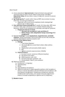
Renal Uworld 1) Acute, symptomatic hypernatremia ( impaired mental status/seizure) requires prompt increase in serum sodium concentration with 3% or Hypertonic Saline. No more than a rate of .5 mEq/L/hr to avoid cns osmotic demylination 2) IgA Nephropathy usually within 5 days of URTI, more common in young adult men, recurrent gross hematuria a. Diagnosed with normal serum complement levels, mesangial IgA deposits seen in kidney biopsy 3) Post infectious Glomerulonephritis usually 10-21 days after URTI, more common in kids ( age 6-10) but can occur in adults. Presents with gross hematuria. Adults can be asymptomatic or develop acute nephritic syndrome a. Diagnosed with Low C3 levels i. Elevated anti-stretolysin O and anti-DNAse B ii. Kidney biopsy with subepithelial humps consisting of C3 complement 4) Repeated vomiting causes hypokalemic, hypochloremic, metabolic alkalosis. Volume resuscitation with normal saline corrects the contraction alkalosis. Hypokalemia should be treated as well. 5) Prerenal Acute Kidney Injury a. Etiology: i. True Volume Depletion ii. Decreased effective arterial blood volume ( Heart failure, cirrhosis) iii. Displacement of intravasulcar fluids ( sepsis) iv. RAS v. Afferent arteriole vasoconstriction ( NSAIDs) b. Clinical Features: i. Increase in serum creatinine ( 50% from baseline) ii. Decreased urine output iii. BUN/Creatinine ratio >20:1 iv. Fractional Excretion of Sodium 3-3.5g/day) b. Hypoalbumenia c. Edema d. Hyperlipidemia and Lipiduria e. Nephrotic syndrome is a hypercoaguable condition which manifests as venous or arterial thrombosis, and even PE. Renal vein thrombosis is the most frequent manifestation. Complications of nephrotic syndrome include protein malnutrition, iron resisiten microcytic hypochromic anemia, increased susceptibility to infection and vit D deficiency 7) Bladder Cancer
a. Bladder tumors are the most common malignancy associated with painless hematuria in adults. b. Initial assessment of painless gross hematuria includes urine analysis to rule out UTI and confirm micro hematuria. c. Risk factors include age greater than 35, smoking history, occupation history (chemicals dyes) and drug exposure (cyclophosphamide). d. Patient should have imaging (Contrast CT Scan) and cystoscopy to evaluate the bladder and urethra. 8) Uric Acid Stone a. Tx of uric acid stones includes hydration, alkalinization of the urine, and low purine diet. Alkalinization of the urine to pH 6-6.5 with oral potassium citrate is recommeneded as urinc acid stones are highly soluable in alkaline urine. 9) Casts a. Muddy Brown Granular Casts Acute Tubular Necrosis b. RBC Casts Glomerulonephritis c. WBC Casts Interstial Nephritis and Pyelonephritis d. Fatty Casts Nephrotic Syndrome e. Broad and Waxy Casts Chronic Renal Failure 10)
Management of Hypercalcemia a. Severe Hypercalcemia ( calcium >14 ) or Symptomatic i. Aggressive Saline Hydration ( Normal Saline) to restore intravascular volume and promote urinary calcium excretion ii. Calcitonin – inhibits osteoclast mediated bone resorption, quickly reduces calcium concentration b. Moderate Hypercalcemia ( 12-14) i. No immediate treatment required unless symptomatic, then same treatment as severe hypercalcemia 11) SLE a. Photosensitive skin ( sun burn) b. Thrombocytopenia c. Glomerulopnephritis ( renal failure with erythrocyte casts, proteinuria, hyptertension) w low complememnt levels ( C3, C4) d. Positive ANA, anti double stranded DNA, and anti Smith 12)Uncomplicated cystitis commonly occurs in healthy patients and has a low risk of treatment failure. Urinalysis confirms the diagnosis. Patients can be treated without a urine culture, which may be done later in those who fail initial therapy. Oral TMP/SMX, Nitrofurantoin, and fosfomycin are effective first line treatment options. 13)Complicated Cystitis associated with factors that increase risk of antibiotic resistence or tx failure ( diabetes, catheters, pregnancy, urinary tract obstruction). These patient should have URINE CULTURE prior to therapy. Tx: Flouoroquinolones, extended spectrum antibiotic ( ampicillin/gentamicin) for more severe cases
14)Pyelonephritis require culture prior to initiating treatment. Stable patients with uncomplicated pyelonephritis can be treated with oral antibiotics, usually a floroquinolone ( ciprofloxacin, levofloxacin), but unstable patients and those with complicated infection require IV antibiotics ( ceftriaxone) 15)Uric Acid stones, which are radiolucent, have to be evaluated by CT of the abdomen, ultrasonography, or IV pyelography 16) Loop Diuretics are frequently administered to cirrhotic patients, with volume overload and ascites. Potential side effects include hypokalemia, metabolic alkalsosis, and prerenal kidney injury 17) Primary Renal Causes of Nephrotic Syndrome a. Focal Segmental Glomerulosclerosis African American, Hispanic, Obesity, HIV/Heroin Use b. Membranous Nephropathy Adenocarcinoma ( breast and lung) , NSAIDs, Hep B, SLE c. Membranoproliferative Glomerulonephritis Hep C, Hep B, Lipodystrophy d. Minimal Change Disease Nsaids, HODGKINS LYMPHOMA* e. IgA Nephropathy Upper Resp Infection 18)0.9% IV Normal Saline is preferred for treating hypovolemic hypernatremia. The fluid can be switched to a hypotonic fluid ( 5% Dextrose) for free water supplementation once the patient in euvolemic 19)ACE Inhibitor or ARB are indicated for initial therapy in patients with hypertension and renal artery stenosis. Renal artery stenting or surgical revascularization is reserved for patients with resistant htn or recurrent flash pulmonary edema or refractory Heart Failure due to severe htn. 20) Patients with nephrotic syndrome have increased risked for atherosclerosis ( due to hyperlipidemia) and arteriovenous thrombosis ( due to loss of antithrombin III). Atherosclerosis and hypercoagulability increase the risk for stroke and MI. 21) Aspirin Intoxication Mixed Resp Alkalosis and Metabolic Acidosis 22) SIADH a. Etiologies i. Medications ( carbamazepine, SSRI, NSAIDs) ii. Lung Disease ( pneumonia) iii. Ectopic ADH Secretion ( Small Cell Lung Cancer) b. Clinical Features i. Mild/moderate hyponatremia- nausea, forgetfulness ii. Severe hyponatremia-seizure coma iii. Euvolemia ( moist membranes, no edema, no JVD) c. Laboratory Findings i. Hyponatremia ii. Serum Osmolality 100 iv. Urine Sodium >40 d. Management i. Fluid Restriction +/- salt tablets
ii. Hypertonic (3%) Saline for severe hyponatremia iii. Patients with SIADH who are asymptomatic or have mild symptoms usually respond to fluid restriction. Patient who have severe symptoms require treatment with HYPERTONIC 3% SALINE 23) Oxalate absorption is increased in Crohn Disease and other intestinal diseases causing fat malabsorption. Increased absorption is the most common cause of symptomatic hyperoxaluria and oxalate stone formation 24) NEPHROLITHIASIS MANAGEMENT a. Imaging Study CT scan of the abdomen without contrast is the investigation of choice because of its high sensitivity and specificity. It has the advantage over x-ray (KUB) in detecting radiolucent stones b. Narcotics and NSAIDs pain relief c. Size of the stone Stones measuring less than 5mm in diameter typically pass spontaneously with conservative treatment. This includes a fluid intake of greatrer than 2L daily. Increased hydration increases uriniary flow rate and lower urinary solute concentration thus preventing stone formation d. You don’t need a metabolic evaluation when a patient presents with his first stone. In patients with recurrent stone, 24 hr urine is collected to identify any underlying metabolic disorder 25) Collapsing focal and segmental glomerulosclerosis is the most common form of glomerulopathy associated with HIV. Typical presentation of focal segmental glomerulosclerosis includes nephrotic range proteinuria, azotemia and normal sized kidneys 26) Type 4 renal tubular acidosis ( Hyperkalemic Renal Tubular Acidosis) is characterized by non anion gap metabolic acidosis, persistent hyperkalemia, and mild to moderate renal insufficiency. Commonly occurs in patients with poorly controlled diabetes. 27)Most common kidney stone- calcium oxalate 28) Acute Interstitial Nephritis a. Causes: Drugs ( Penicillins, TMP-SMX, Rifampin, Cephalosporins, NSAIDs) b. Clinical Features: i. Maculopapular rash ii. Fever iii. New drug exposure iv. +/- Arthralgia c. Lab Findings: i. Acute Kidney Injury ii. Pyuria, Heamturia, WBC Casts iii. Eosinophilia, Urinary Eosinophils iv. Renal Biopsy: Inflammatory Infiltrate, Edema d. Management: i. Discontinue Drug ii. +/- Systemic Glucocorticoids
29)Glomerular Hyperfiltration is the earliest renal abnormality seen in diabetic nephropathy. It is also the major pathophysiological mechanism of glomerular injury in these patients. Thickening of the glomerular basement membrane is the first change that can be quantitated 30) Hyperkalemia is a medical emergency. Therapy involves 3 goals: Stabilizing the cardiac membrane ( calcium chloride or calcium gluconate), shifting potassium intracellulary, and decreasing total body potassium content. Insulin/glucose administration is the quickest way to lower serum concentration 31) Renal Vein Thrombosis is most commonly associated with membranous glomerulopathy. ( Loss of Antithrombin III) 32) Ureteral calculi may cause flank or abdominal pain radiating to the perineum often with nausea and vomiting. Ultrasound or a non contrast CT scna of the abdomen and pelvis are the imaging modalities of choice to fconfirm the diagnosis. Ultrasonography is preferred in pregnant patients to reduce radiation exposure 33) CT scan of the abdomen is the most sensitive and specific test for diagnosing Renal Cell Carcinoma
UWORLD STEP 3: Nephro: 1) Urethral diverticulum is suspected in patients with dysuria, postvoid dribbling, dyspareunia and an anterior vaginal wall mass on pelvic examination. Dx confirmed with Pelvic MRI or Transvaginal US. Treatment options include manual decompression, needle aspiration, or surgical repair. 2) Methylene blue- instilled into bladder to diagnose urinary incontence due to Vesicovaginal Fistula. 3) Posterior urethral valves- antenatal ultrasound will show b/l hydronephrosis, thickened and dilated bladder and a dilated proximal urethra. If the obstruction is sevrre, oligohydramnios can result and lead to POTTER sequencepulmonary hypoplasia and flattened facies. Dx via a voiding cystourethrogram. ( voiding cystourethrogram- can dx vesicocourteral reflux and posterior urethral valves). Once PUV is confirmed infants should have a foley catheter placed to relieve obstruction. When stablaized, cystoscopy allows direct visulation and ablation of the valve. 4) Normal serum compliments and onset of symptoms days after an URI are suggestive of IGA Nephropathy. IgA Immune complex deposition in the mesangium. Flank pain is common due to stretching of the renal capsule. Recurrent episodes of hematuria are common but IgA nephropathy is usually benign disease with no chronic kidney injury. 5) In non pregnant patients uncomplicated cystitis can be treated empirically. A urine culture is indicated if symptoms do not respond to antibiotics. In pregnancy for acute cystitis use – cephalexin/Fosfomycin/amoxicillinclavulonate. Gentamicin can cause congenital deafness.
6) CT scan of abd is used to dx renal cell carcinoma. If tumor localized to the kidney, nephrectomy is curative. If renal mass is confined within the capsule partial nephrectomy can be offered. 7) Vigorous hematopoeisis occurs with admin of Erythropoeisis stimulating agents and can lead to rapid depletion of iron stores; patients should have iron levels checked 8) No indications to do a genetic test to screen for ADPKD if patient has multiple renal cyst on ultra sound. ADPKD presents age 30-40 /flank pain/hematuria/htn/palpable abd masses ( often b/l) . Extrarenal features: cerebral aneurysm/hepatic+pancreatic cyst/mvp/aortic regurg/colonic diverticulosis/ventral/inguinal hernia . Control BP with ACE inhibitor. Add statin to reduce CV risk. 9) Patients greater than the age of 18 w/ a family hx of ADPKD should be screened with renal ultrasound and should receive counseling. Counseling should be done prior to screening as a positive test can have psychological/insurability/and employment ramifications. 10) Kidney biopsy is indicated prior to tx initiation to guide therapy in all patients with sle w/ significant renal involvement. Anti double stranded DNA and complement levels can be used to monitor for active renal involvement in patients with SLE. Antinuclear antibody titers do not correlate with renal disease activity. 11) IV NS or Sodium bicarb before and after contrast to prevent contrast induced nephropathy 12)...
Similar Free PDFs

Renal Uworld And Thats All
- 6 Pages

Uworld-adult-urinary renal-1
- 17 Pages

Nclex Charts and uworld prep
- 38 Pages

CPAT3202 Renal and Intestinal
- 36 Pages

Renal
- 10 Pages

UWorld Medical Surgical Nursing
- 142 Pages

Uworld Maternity - Questions
- 96 Pages

Uworld-pediatrics study notes
- 53 Pages

Uworld NCLEX Notes
- 40 Pages

All and Also All: Synthesis
- 1 Pages

UWorld Critical Care
- 14 Pages

Uworld adult infectious 2
- 10 Pages

UWorld Peds 9
- 100 Pages

Uworld-adult-visual auditory-1
- 5 Pages
Popular Institutions
- Tinajero National High School - Annex
- Politeknik Caltex Riau
- Yokohama City University
- SGT University
- University of Al-Qadisiyah
- Divine Word College of Vigan
- Techniek College Rotterdam
- Universidade de Santiago
- Universiti Teknologi MARA Cawangan Johor Kampus Pasir Gudang
- Poltekkes Kemenkes Yogyakarta
- Baguio City National High School
- Colegio san marcos
- preparatoria uno
- Centro de Bachillerato Tecnológico Industrial y de Servicios No. 107
- Dalian Maritime University
- Quang Trung Secondary School
- Colegio Tecnológico en Informática
- Corporación Regional de Educación Superior
- Grupo CEDVA
- Dar Al Uloom University
- Centro de Estudios Preuniversitarios de la Universidad Nacional de Ingeniería
- 上智大学
- Aakash International School, Nuna Majara
- San Felipe Neri Catholic School
- Kang Chiao International School - New Taipei City
- Misamis Occidental National High School
- Institución Educativa Escuela Normal Juan Ladrilleros
- Kolehiyo ng Pantukan
- Batanes State College
- Instituto Continental
- Sekolah Menengah Kejuruan Kesehatan Kaltara (Tarakan)
- Colegio de La Inmaculada Concepcion - Cebu

