Table of lower limb muscles PDF

| Title | Table of lower limb muscles |
|---|---|
| Course | Anatomy, Histology and Embryology 1 |
| Institution | Debreceni Egyetem |
| Pages | 7 |
| File Size | 300.9 KB |
| File Type | |
| Total Downloads | 119 |
| Total Views | 199 |
Summary
A GUIDE TO THE MUSCLES OF THE LOWER LIMB Category Muscle Function Origin Insertion Nerve Blood Supply Gluteal Region Gluteus maximus Hip extensor Lateral rotator (H) Suface of ilium, sacrum Inferior gluteal (L5, S1, S2) Inf. Sup. Gluteal Gluteus medius Hip abductor Medial rotator (H) Hip abductor Me...
Description
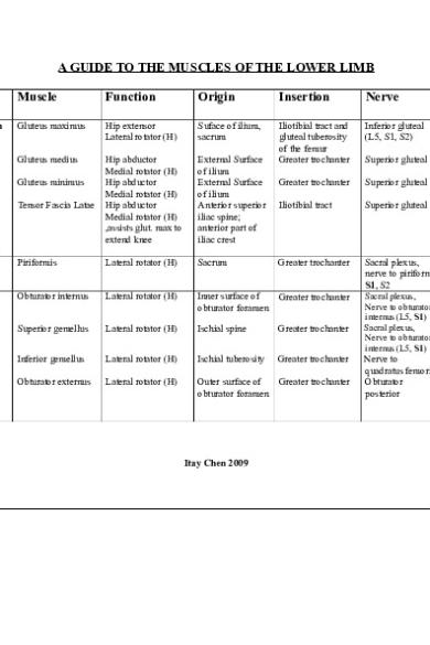
A GUIDE TO THE MUSCLES OF THE LOWER LIMB Category
Muscle
Function
Origin
Insertion
Nerve
Blood Supply
Gluteal Region
Gluteus maximus
Hip extensor Lateral rotator (H)
Suface of ilium, sacrum
Inferior gluteal (L5, S1, S2)
Inf. & Sup. Gluteal
Gluteus medius
Hip abductor Medial rotator (H) Hip abductor Medial rotator (H) Hip abductor Medial rotator (H) ,assists glut. max to extend knee
External Surface of ilium External Surface of ilium Anterior superior iliac spine; anterior part of iliac crest
Iliotibial tract and gluteal tuberosity of the femur Greater trochanter
Superior gluteal
Superior gluteal
Greater trochanter
Superior gluteal
Superior gluteal
Iliotibial tract
Superior gluteal
Superior gluteal
Piriformis
Lateral rotator (H)
Sacrum
Greater trochanter
Sacral plexus, nerve to piriformis S1, S2
Inf. & Sup. Gluteal
Obturator internus
Lateral rotator (H)
Inner surface of obturator foramen
Greater trochanter
Inf. Gluteal
Superior gemellus
Lateral rotator (H)
Ischial spine
Greater trochanter
Sacral plexus, Nerve to obturator internus (L5, S1) Sacral plexus, Nerve to obturator internus (L5, S1)
Inferior gemellus
Lateral rotator (H)
Ischial tuberosity
Greater trochanter
Obturator externus
Lateral rotator (H)
Outer surface of obturator foramen
Greater trochanter
Gluteus minimus Tensor Fascia Latae
Pelvic Region
Itay Chen 2009
Nerve to quadratus femoris Obturator posterior
Inf. Gluteal Inf. Gluteal Med. circumflex femoral & Obturator
Hip exceptions
Quadratus femoris
Lateral rotator (H)
ischial tuberosity
intertrochanteric crest of femur
Nerve to quadratus femoris
Inferior glutial artery
Iliacus (part of iliopsoas)
flexing thigh at hip joint
Iliac crest, iliac fossa, ala of sacrum, and anterior sacroiliac ligaments Sides of T12-L5 vertebrae and discs between them. Sides of T12-L1 vertebrae and intervertebral disc superior Pubic ramus
Tendon of psoas major, lesser trochanter.
Femoral nerve
Medial femoral circumflex
Lesser trochanter of femur
Anterior rami of lumbar nerves
Lumbar branch of iliolumbar artery
Pectineal line, iliopectineal eminence. Pectineal line of femur
Anterior rami of lumbar nerves
lumbar artery
Femoral
Anterior superior iliac spine
Upper medial tibia
Femoral
Med. Circumflex femoral & Obturator Femoral
Anterior inferior iliac spine and ilium superior to acetabulum Intertrochanteric line and medial lip of linea aspera of femur Greater trochanter and lateral lip of
Patellar ligament
Femoral
Lateral circumflex femoral
Patellar ligament
Femoral
Femoral (profunda femoris)
Patellar ligament
Femoral
Lateral circumflex
Psoas major (part of flexing thigh at hip iliopasoas) joint
Anterior Thigh
Psoas minor
flexing thigh at hip joint
Pectineus
Hip Flexor Hip Adductor Medial roatation
Sartorius
Hip Flexor Hip Abductor Lateral Rotator (H) Knee extensor Extend leg at knee joint, help in hip flexion.
Rectus femoris
Vastus medialis
Extend leg at knee joint
Vastus lateralis
Extend leg at knee joint
Itay Chen 2009
Medial Thigh
Vastus intermedius
Extend leg at knee joint
Gracilis
Knee Flexor Hip Adductor
Adductor magnus
Adductor part: adduction Hamstrings part: extends thigh
Posterior Thigh
linea aspera of femur Anterior and lateral surfaces of shaft of femur
femoral Patellar ligament
Femoral
Lateral circumflex femoral
Inferior Pubic ramus
Upper medial tibia
Obturator
Adductor part: inferior ramus of pubis, ramus of ischium Hamstrings part: ischial tuberosity
Adductor part: gluteal tuberosity, linea aspera, medial supracondylar line Hamstrings part: adductor tubercle of femur Pectineal line and proximal part of linea aspera of femur Middle third of linea aspera of femur
Adductor part: obturator nerve,
Med circumflex femoral & obturator Med circumflex femoral & obturator
Adductor brevis
Hip Adductor Thigh flexor
Pubic ramus
Adductor Longus
Hip Adductor Thigh flexion
Anterior surface of body of pubis
Semitendinosus
Hip Extensor Knee Flexor (roatat the leg medialy
Ischial tuberosity
Itay Chen 2009
Medial surface of superior part of tibia
Hamstrings part: tibial part of sciatic nerve.
Obturator
Med circumflex femoral & obturator
Obturator
Med circumflex femoral & obturator
Tibial
Perforating br. Of deep femoral
when the knee is flexed) Semimembranosus Hip Extensor Knee Flexor (roatat the leg medialy when the knee is flexed) Long head of biceps Hip Extensor femoris Knee Flexor Lateral rotator (H) Short head of Knee Flexor biceps femoris Lateral rotator (H)
Posterior Leg
Gastrocnemius
Plantar flexion(A) Knee Flexor
Plantaris
Plantar flexion(A)
Soleus
Plantar flexion(A)
Flexor digitorum longus
Plantar flexion(A) Flexes toes 2-5, supports longitudinal arches of foot
Flexor hallucis longus
Plantar flexion(A) Flexes toe 1, supports medial
Ischial tuberosity
Posterior part of medial condyle of tibia
Tibial
Perforating br. Of deep femoral
Ischial tuberosity
Fibular head
Tibial
Perforating br. Of deep femoral
linea aspera and lateral supracondylar line of femur Medial and lateral epicondyles of femur lateral supracondylar line of distal femur Posterior aspect of head of fibula, medial border of tibial shaft, Posterior surface of tibia
Fibular head
Common Fibular
Perforating br. Of deep femoral
Calcaneus (via calcaneal tendon) Calcaneus (via calcaneal tendon)
Tibial
Sural br. Of popliteus
Tibial
Sural br of popliteus
Calcaneus (via calcaneal tendon)
Tibial
Sural br of popliteus & posterior tibial
bases of 2nd - 5th distal phalanges (go under medial malleulus)
Tibial
Posterior tibial
Base of distal phalanx of great toe (go under
Tibial
Posterior tibial
posterior surface of fibula, interosseous Itay Chen 2009
Tibialis posterior
Dorsum of Foot
Lateral Leg
Anterior Leg
longitudinal arches of foot Plantar flexion(A) Inversion
Popliteus
Rotates knee medially and flexes the leg on the thigh
Extensor digitorum brevis
Extensor of toes (by aiding the extensor digitorum longus)
Extensor Hallucis Brevis
Extends great toe
Fibularis longus
Plantarflexion (A) Eversion
Fibularis brevis
Plantarflexion (A) Eversion
Tibialis anterior
Dorsiflexes ankle and inverts foot
membrane
medial malleulus)
Posterior surface of tibia and fibula &interosseous membrane
Navicular, cuniform, cuboid, bases of 2-4 metatarsals. (go under medial malleulus) Posterior surface of tibia, superior to soleal line
Lateral surface of lateral condyle of femur and lateral meniscus Calcaeneus, interosseous talocalcaneal ligament In common with extensor digitorum brevis Head and superior two thirds of lateral surface of fibula
Long flexor tendons of four medial toes
Dorsal aspect of base of proximal phalanx 1 Metatarsal of toe #1 & medial cuneiform (go under lateral malleulus) Inferior two thirds Base Metatarsal of of lateral surface toe #5 (go under of fibula lateral malleulus) Lateral condyle Medial and superior cuneiform/first lateral surface of metatarsal tibia and interosseous membrane
Itay Chen 2009
Tibial
Posterior tibial, peroneal
Tibial nerve
Medial inferior genicular & Posterior tibial
Deep fibular
Dorsalis pedis
Deep fibular
Dorsalis pedis
Superficial fibular
Fibular artery
Superficial fibular
Fibular artery
Deep fibular
Anterior tibial
Plantar Foot Level 1
Extensor hallucis longus
Extends great toe and dorsiflexes ankle
Extensor digitorum longus
Dorsiflexion (A) Extensor of toes
Fibularis tertius
Distal phalanx of toe #1 (dorsum)
Deep fibular
Anterior tibial
Extensor expansion of lateral 4 toes
Deep fibular
Anterior tibial
Dorsiflexes ankle and aids in eversion of foot
Middle part of anterior surface of fibula and interosseous membrane Lateral condyle of tibia and medial surface of fibula and interosseous membrane distal anterior surface of the fibula
metatarsal of toe #5 Deep fibular
Anterior tibial
Abductor hallucis
Abducts toe #1 Flexes toe #1
Medial tubercle of calcaeneum
Medial plantar (Tibial)
Medial plantar (post. Tibial)
Flexor digitorum brevis Abductor digiti minimi Quadratus plantae
Flexes lateral four digits Abducts and flexes little toe Assists flexor digitorum longus in flexing lateral four digits Flex proximal phalanges, extend middle and distal phalanges of lateral four digits
Medial tubercle of calcaeneum Calcaeneum
Medial side of base of proximal phalanx of 1st digit Middle phalanx of toes #2-5 Lateral base of phalanx #5 Tendons of flexor digitarum longus
Medial plantar (Tibial) Lateral plantar (Tibial) Lateral plantar (Tibial)
Medial plantar (post. Tibial) Lateral plantar (post. Tibial) Lateral plantar (post. Tibial)
Medial one: medial plantar nerve. Lateral three: lateral plantar nerve.
Medial and Lateral plantar arteries
Level 2
Lumbricals
Calcaeneum
Tendons of flexor digitorum longus
Itay Chen 2009
Medial aspect of expansion over lateral four digits
Flexor digitorum longus tendon
Level 3
Flexor hallucis brevis Adductor hallucis
Passes in the sustentaculum tali and crosses with the flexor hallucis longus tendon Flexes big toe
Cuboid and lateral cuneiform Oblique Head: proximal ends of middle 3 metatarsal bones; Transverse Head: MTP ligaments of middle 3 toes Base of metatarsal bone #5
Adducts big toe and assists in transverse arch of foot.
Flexor digiti minimi Flexes pinky toe brevis
Level 4
Flexor hallucis longus tendon Plantar interossei 3
Dorsal interossei 4
Adduct digits (2-4) and flex metatarsophalangeal joints Abduct digits (2-4) and flex metatarsophalangeal joints
Bases and medial sides of metatarsals 3-5 Adjacent sides of metatarsals
Itay Chen 2009
Base of proximal phalanx of toe #1 Lateral side of proximal phalanx of big toe #1
Medial plantar (Tibial) Lateral plantar
Medial plantar (post. Tibial) lateral plantar (post. Tibial)
base of the proximal phalanx of toe #5
Superficial branch of lateral plantar nerve
Lateral plantar (post. Tibial)
Medial sides of bases of phalanges 3-5
Lateral plantar
Lateral plantar (post. Tibial)
Phlanages and dorsal expansion of corresponding toes
Lateral plantar
Lateral plantar (post. Tibial)...
Similar Free PDFs

Table of lower limb muscles
- 7 Pages

Muscles of Lower Limb - musclee
- 7 Pages

Innervation of Lower limb
- 6 Pages

Muscles of Upper Limb
- 11 Pages

Vasculature of Lower limb
- 5 Pages

Lower Limb Questions
- 6 Pages

table of shoulder muscles
- 2 Pages

Muscles of the lower limb2013
- 46 Pages

Table of Muscles of Mastication
- 1 Pages

Lower Limb mcqs
- 55 Pages

Lower limb notes
- 22 Pages

lower limb anatomy questions
- 4 Pages

Lower limb chart
- 9 Pages

Kenhub lower limb muscle
- 9 Pages

Mnemonics - Lower Limb
- 8 Pages
Popular Institutions
- Tinajero National High School - Annex
- Politeknik Caltex Riau
- Yokohama City University
- SGT University
- University of Al-Qadisiyah
- Divine Word College of Vigan
- Techniek College Rotterdam
- Universidade de Santiago
- Universiti Teknologi MARA Cawangan Johor Kampus Pasir Gudang
- Poltekkes Kemenkes Yogyakarta
- Baguio City National High School
- Colegio san marcos
- preparatoria uno
- Centro de Bachillerato Tecnológico Industrial y de Servicios No. 107
- Dalian Maritime University
- Quang Trung Secondary School
- Colegio Tecnológico en Informática
- Corporación Regional de Educación Superior
- Grupo CEDVA
- Dar Al Uloom University
- Centro de Estudios Preuniversitarios de la Universidad Nacional de Ingeniería
- 上智大学
- Aakash International School, Nuna Majara
- San Felipe Neri Catholic School
- Kang Chiao International School - New Taipei City
- Misamis Occidental National High School
- Institución Educativa Escuela Normal Juan Ladrilleros
- Kolehiyo ng Pantukan
- Batanes State College
- Instituto Continental
- Sekolah Menengah Kejuruan Kesehatan Kaltara (Tarakan)
- Colegio de La Inmaculada Concepcion - Cebu
