Week 4 lab anatomy and physiology assignment PDF

| Title | Week 4 lab anatomy and physiology assignment |
|---|---|
| Author | doris Adebanjo |
| Course | anatomy & physiology 1 |
| Institution | Chamberlain University |
| Pages | 18 |
| File Size | 908.2 KB |
| File Type | |
| Total Views | 125 |
Summary
week 4 lab , anatomy and physiology week 4 assignment document...
Description
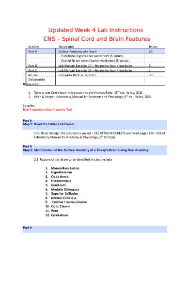
Updated Week 4 Lab Instructions CNS – Spinal Cord and Brain Features Activity Part A
Part B Part C All Lab Deliverables References:
Deliverable Surface Anatomy of a Brain --Functional Significance worksheet (5 points) --Cranial Nerve Identification worksheet (5 points) Lab Manual Exercise 17 – Reviewing Your Knowledge Lab Manual Exercise 18 – Reviewing Your Knowledge Complete Parts A , B, and C
Points 20
5 5 30
1. Tortora and Derrickson Introduction to the Human Body, 15th ed., Wiley, 2016. 2. Allen & Harper, Laboratory Manual for Anatomy and Physiology, 6th ed., Wiley, 2016. Supplies: Real Anatomy online Anatomy Tool Part A Step 1: Read the Entire Lab Packet
1.0– Read through the laboratory packet – SEE ATTACHED SHEETS and read pages 324 - 326 of Laboratory Manual for Anatomy & Physiology (5th Edition) Part A Step 2: Identification of the Surface Anatomy of a Sheep’s Brain Using Real Anatomy
2.3 Regions of the brain to be identified in class include 1. 2. 3. 4. 5. 6. 7. 8. 9. 10. 11. 12.
Part A
Mammillary bodies Hypothalamus Optic Nerve Hippocampus Cerebrum Medulla Oblongata Superior Colliculus Inferior Colliculus Trochlear nucleus/nerve Optic Chiasm Pons Cerebellum
Step 3: Functional Significance Worksheet and Cranial Nerve Identification
3.1 –Complete the functional significance worksheet corresponding to the above regions of the brain 3.2 – Cranial Nerve Identification – Name/Modality/Function –complete the cranial nerve table 3.3 - Submit all completed worksheets directly to your professor or upload to Canvas prior to the beginning of the next scheduled class Part B - Lab Manual Exercise 17: Spinal Cord Structure and Function
From Allen and Harper, Laboratory Manual for Anatomy and Physiology 6th Edition 4.0 – Read background pages 277 – 282. 4.1 –Answer all questions from Laboratory Manual: Reviewing Your Knowledge, pages 283 – 284. 4.2 – Submit all completed worksheets directly to your professor or upload to Canvas prior to the beginning of the next scheduled class Part C - Lab Manual Exercise 18: Spinal Nerves
From Allen and Harper, Laboratory Manual for Anatomy and Physiology 6th Edition 5.0 – Read background pages 287 - 292. 5.1 – Answer all questions from Laboratory Manual: Reviewing Your Knowledge, pages 293 – 296. 5.3 – Submit all completed worksheets directly to your professor or upload to Canvas prior to the beginning of the next scheduled class
W4 Lab Worksheet -- Surface Anatomy of a Sheep’s Brain Note to Students: Print the following pages for use.
IDENTIFY THE FOLLOWING STRUCTURES ON THE WHOLE SHEEP BRAIN – EACH MEMBER OF YOUR GROUP MUST BE PREPARED FOR QUIZZING BY THE INSTRUCTOR CEREBRUM
CEREBELLUM
TROCHLEAR NERVE HYPOTHALAMUS
SUPERIOR FRONTAL SULCUS INFUNDIBULUM
OPTIC TRACT
LATERAL OLFACTORY STRIA
LONGITUDINAL FISSURE
PONS
MEDULLA OBLONGATA SUPERIOR COLLICULUS OLFACTORY BULB MEDIAL OLFACTORY STRIA
LONGITUTIDNA L FISSURE INFERIOR COLLICULUS OPTIC NERVE RHINAL FISSURE
CRUCIATE FISSURE MAMMILARY BODIES OPTIC CHIASM HIPPOCAMPAL GYRUS
Functional Significance of Structures Name: ____________doris Adebanjo_________ Describe the functional significance of the following structures: Cerebellum Mammillary bodies Hypothalamus
Optic Nerve Hippocampus
Regulates our posture and balance, coordinates skeletal muscle movements Functions as relay stations for reflexes related to the sense of smell Regulates homeostasis, hormonal functions and controls pituitary gland, emotions, temperature eating and drinking. Conducts nerve impulses for vision, sensory.
Superior Colliculus
Consists of cells capable of mitosis, responsible for some parts of memory. Intelligence, communication, learning, memory, reasoning , emotions, interprets sensory input and initiates skeletal muscle contraction Regulates heartbeat, blood vessel diameter, breathing, provides cerebellum with instructions when learning new motor skills. Coordinates vomiting, swallowing, sneezing, coughing. Visual reflex
Inferior Colliculus
Auditory reflex
Trochlear nucleus/nerve:
Provides motor impulses that control movement of Vital to vision, left and right optic nerves intersect at the chiasm thereby creating the hallmark xshape also enables eye-hand coordination Connects upper and lower parts of the brain by serving as a message station. Relays messages from the cortex and the cerebellum. Involved in control of breathing, sensations of hearing, taste and balance.
Cerebrum
Medulla Oblongata
Optic Chiasm
Pons
Complete the following table for the twelve cranial nerves that you are identifying on the sheep brain (see last page of dissection guide)
Cranial Nerve Table Number I
Name Olfactory
Sensory/Motor/Mixed sensory
Function(s) smell
II
optic
sensory
sight
III
Oculomotor
motor
Eye movement, pupil constriction
IV
Trochlear
motor
Eyeball movement, proprioception
V
Trigeminal ophthalmic/maxillary./mandibular branch
mixed
VI
Abducens
motor
Sensations of touch and pain from facial skin, nose, mouth and tongue, proprioception motor control of chewing Eyeball movement, proprioception.
VII
Facial
mixed
VIII
Vestibulocochlear: vestibular /cochlear branch
sensory
IX
Glossopharyngeal
mixed
X
Vagus
mixed
XI
Accessory
motor
Movement if facial muscles, tear and saliva secretion, sense of taste and proprioception Sense of equilibrium and hearing Taste, touch, pain from tongue and pharynx, chemoreceptors that monitor o2 and co2. Blood pressure receptors, movements of tongue and swallowing, secretion of saliva. Taste, voice box, co2/o2 in blood, swallowing, ! HR Head movement
XII
Hypoglossal
motor
Speech and swallowing
Part B Reviewing Your Knowledge A) 1. Endoneurium 2 . perineurium 3 epineurium B) 1. Posterior ramus 2 rami comunicantes 3 anterior ramus C) 1 lumbar 2 sacral 3 cervical 4 brachial 5 brachial 6 sacral 7 lumbar 8 brachial 9 sacral 10 sacral D) 1 tibial 2 common fibular 3 31 4 tibial 51 6 motor 7 12 8 obturator 95 10 5 11 sensory 12 8 13 axillary 14 radial 15 musculotaneous 16 phrenic 17 ulnar
18
E
1 spinal nerve 2 epimeurium 3 perineurium 4 fassicle 5 endoneurium 6 axon
Figure 18.7 1 axillary nerve 2 musculocutaneous nerve 3 median nerve 4 ulnar nerve 5radial nerve 6 phrenic nerve Figure 18.8 1 femoral nerve 2 3 obturator nerve 4 sciatic nerve 5 common fibular nerve 6 femoral cutaneous nerve
Using Your Knowledge 1 median nerve
2 radial nerve 3 sciatic nerve 4 ulnar nerve 5 c8 6 obturator nerve 7 common fibular nerve 8 sciatic nerve 9 axillary nerve 10 ulnar nerve
Using Real Anatomy: Identification of Brain Structures Accessing Real Anatomy: Open ModulesWeek 4 Onsite lab and click the Real Anatomy link.
For Identification Topics 1-6, Please use the following tab in Real Anatomy: After opening the “Nervous” tab in Real Anatomy, select Brain. Hover your mouse over the “Brain, right lateral view” to see all the views available. Explore each following along with the structures described below.
For Identification Topics 7-10, Please use the following tab in Real Anatomy: Type “Cranial Nerves” in the search bar above. You should then be able to select “Inferior view of the brain and cranial nerves”.
Brain Dissection Instructions 1. Major Subdivisions It is important to be able to recognize the major subdivisions of the brain ‘at a glance.’ As you look at your sheep brain you need to recognize these subdivisions from a variety of perspectives. The first objective is look understand its orientation in a sagittal perspective. The image below shows the left side of the brain with the brain ‘looking’ to the left. The cerebrum, which constitutes the majority of the front of the brain, is to the left. The cerebellum is tucked in behind it and the brainstem (medulla) can be seen sticking out to the right ventral to the cerebellum. Whenever you look at a brain you should be able to quickly orient yourself by finding these three subdivisions. 2.
Top view
This is the dorsal view of the brain (the top, looking down). Again, the brain is facing to the left. The cerebrum and cerebellum are easily seen. The ‘tail’ end of the medulla is also visible to the right. A word of caution – you may become dependent on seeing the brain ‘looking left.’ With your brain (the one in the tray), you should practice looking at every brain structure from all directions – spin and rotate the brain tissue to develop the 3-dimensional perspective of the organization of the brain
3.
Ventral View of Brain
This is the bottom of the brain (i.e. the ventral view). The brain stem – including the medulla, pons, and hypothalamus, forms the stalk to which the other parts of the brain attach. The cerebrum can be easily seen wrapping around from the sides going over the top. Only the outside edges of the cerebellum can be seen here – but on closer examination you can see fibers running from the pons that attach it to the cerebellum.
4. Axes Now we will look at the brain again from the same three perspectives and apply the common anatomical terms for referring to direction in ‘brain navigation’. Rostral means toward the front or nose of the animal. Moving rostrally would mean moving to the left in this picture. A synonym for rostral in animals would be anterior in humans as it relates to the brain (please realize that the brain of the sheep in terms of its axis is turned 90 degrees to much of the human anatomical directions). Caudal means towards the tail of the animal. So moving caudally would mean towards the right in this illustration. You can also look ‘dorsally’ and ‘ventrally’ in this image. Moving dorsally would mean moving toward the back of
the animal (which in a human would be posterior rather than dorsal). The dorsal surface of the brain is at the top of this picture. 5. Here, you are introduced to the directional terms ‘medial’ and ‘lateral’. As always, the front of the brain is facing toward the left of the paper. The center of the brain is considered the most medial portion and the sides of the brain are considered the most lateral portions. These terms are used on a continuum and are relative to each other. As you move toward the midline (midsagittal) plane of the brain, you are moving medially.
6. The longitudinal fissure divides the left and right cerebral hemispheres along the midline. If you look at the rostral pole of each hemisphere you can see the superior frontal sulcus. This sulcus ‘T’s into the cruciate fissure. Why is one a ‘sulcus’ and the other is a ‘fissure’? TRADITION IS THE REAL REASON!!!. You should also be able to pick out the following lobes on your brain – occipital, temporal, parietal and frontal – look in your book and relate it to the sheep brain (the cruciate sulcus is the equivalent of the central fissure in man).
Remember to Switch Your View In Real Anatomy 7. Midbrain Surface Structures Here the brain has been bent at the rostral and caudal poles to expose the area between the cerebral and cerebellar cortices. You can clearly see the dorsal surface of the midbrain. The two large sets of bumps are called the quadrigeminal complex (4’some). The two large rostral bumps are the superior colliculi (hills). The superior colliculus are located ‘above’ (or in other words superior) to the inferior colliculi along the midbrain surface that are the smaller set of bumps. In the crease just caudal to the inferior colliculus on either side of the midbrain you should be able to see a small whitish nub / fiber band. The trochlear nerve (IV) exits the midbrain here. This is the only cranial nerve that exits from the dorsal surface of the brain. All the other cranial nerves exit from the lateral or ventral aspects of the brain. 8.
Ventral Structures
Your brain may still have some membranes on it and many cranial nerves still attached to the brain (I hope). I’m going to cheat a little at this point and show you a brain that has been stripped clean to allow you to see what is hidden by the thin veil of, the dura, arachnoid and pia maters that still covers much of the brain. Identify: The olfactory bulbs (I) – these are very large in this species (bigger than yours by far) The bright white ‘X’ formed by the optic nerve (II), optic chiasm and optic tract stands out very clearly. The large white cerebral peduncles can be seen running along the ventral surface of the midbrain starting just caudally to the optic tracts. Sometimes students have a hard time seeing them because they are sooooo large. The pons is the bulge of white matter that clearly lifts up from the caudal end of the cerebral peduncles and attach
the cerebellum to the midbrain. The medulla (or as you know it, medulla oblongata) extends caudally from the pons in a tapering fashion until it meets the spinal cord. This image ends with a cut through the rostral spinal cord – if the brainstem is still tapering in a ‘V’ then your cut is through the medulla – it is only when the lateral sides of the brainstem are parallel that you are at the level of the spinal cord. 9. With a slightly closer look at the ventral surface we can now see a few more specific structures: The lateral olfactory stria are easily seen If you look closely you may be able to pick out the medial olfactory stria. The rhinal fissure runs laterally along most of the cerebral cortex. You can also see that it curves onto the medial face of the cerebral hemispheres. Lateral to the optic tracts you can see a fairly smooth section of gray matter that is bounded laterally by the rhinal fissure. This is the hippocampal gyrus. Part of the hippocampus is buried in this part of the temporal lobe as well as the amygdala. The amygdala is located near the rostral tip of the hippocampal gyrus and the hippocampus starts its curving “C”-shaped ram’s horn journey a bit behind the amygdala and follows a parallel path to that of the rhinal fissure. 10. More ventral structures: Looking medially: The longitudinal fissure continues on the ventral surface and divides the two hemispheres rostral to the optic chiasm. Just caudal to the optic chiasm you can see a hole on the midline that also falls about in the middle third of the hypothalamus. You are looking dorsally into the third ventricle through this hole, which was formed when the sheep brain was removed from the skull and the very fragile pituitary and infundibular stalk remained attached to the skull case when the rest of the brain was removed. Remember our journey through the endocrine system and the hypothalamo-pituitary axis. At the caudal end of the hypothalamus there is a bulge. In many sheep brains this bulge appears as a single bump
– however in the human brain it is two distinct bulges – these bulges reminded the old anatomists of the breast buds in developing women (they really needed to get out of the morgue more often) and are called the mammillary bodies. Caudal to the mammillary bodies and between the large cerebral peduncles is a substantial indentation called the interpeduncular cistern (a cistern is a container used to store water or other liquids and was sometimes a covered depression in the earth). This just goes to show that the old neuroanatomists weren’t real creative with their names and just named them after everyday objects – they just sound cool because you get to say them in Latin/Greek. The base of the brain contains the majority of the cranial nerves (we just looked at the one cranial nerve that originates on the dorsal surface – but then exits on the ventral surface – which one was it?)...
Similar Free PDFs

Chicken Anatomy AND Physiology
- 24 Pages

reflex physiology and anatomy
- 4 Pages

Anatomy and Physiology
- 4 Pages

Anatomy-and-physiology
- 197 Pages

Anatomy and Physiology
- 7 Pages

Anatomy and Physiology
- 7 Pages

heart anatomy and physiology
- 4 Pages

anatomy and physiology
- 31 Pages

Anatomy and Physiology notes
- 3 Pages

Chapter 29 anatomy and physiology
- 10 Pages
Popular Institutions
- Tinajero National High School - Annex
- Politeknik Caltex Riau
- Yokohama City University
- SGT University
- University of Al-Qadisiyah
- Divine Word College of Vigan
- Techniek College Rotterdam
- Universidade de Santiago
- Universiti Teknologi MARA Cawangan Johor Kampus Pasir Gudang
- Poltekkes Kemenkes Yogyakarta
- Baguio City National High School
- Colegio san marcos
- preparatoria uno
- Centro de Bachillerato Tecnológico Industrial y de Servicios No. 107
- Dalian Maritime University
- Quang Trung Secondary School
- Colegio Tecnológico en Informática
- Corporación Regional de Educación Superior
- Grupo CEDVA
- Dar Al Uloom University
- Centro de Estudios Preuniversitarios de la Universidad Nacional de Ingeniería
- 上智大学
- Aakash International School, Nuna Majara
- San Felipe Neri Catholic School
- Kang Chiao International School - New Taipei City
- Misamis Occidental National High School
- Institución Educativa Escuela Normal Juan Ladrilleros
- Kolehiyo ng Pantukan
- Batanes State College
- Instituto Continental
- Sekolah Menengah Kejuruan Kesehatan Kaltara (Tarakan)
- Colegio de La Inmaculada Concepcion - Cebu


