Fixation summary notes PDF

| Title | Fixation summary notes |
|---|---|
| Course | Microanatomy and Histotechnology |
| Institution | University of Ontario Institute of Technology |
| Pages | 9 |
| File Size | 236.1 KB |
| File Type | |
| Total Downloads | 264 |
| Total Views | 643 |
Summary
Lecture 1 Class Notes - TISSUE FIXATIONRationale: Fixation is the most important step through the process of histology. Here is where everything starts. A well fixed tissue is the key for a good slide and therefore a good interpretation for diagnosis.Objectives: a) Describe the various fixatives and...
Description
MLSC 3230 Histotechnology & Microanatomy
1
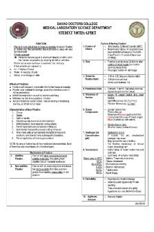
Lecture 1 Class Notes - TISSUE FIXATION Rationale: Fixation is the most important step through the process of histology. Here is where everything starts. A well fixed tissue is the key for a good slide and therefore a good interpretation for diagnosis. Objectives: a) Describe the various fixatives and their uses. b) Learn the difference between autolysis and putrefaction c) Differentiate the fixatives that could impact the final results. d) Learn the chemicals and reagents used in each fixative (refer to the summary fixative chart).
Purpose of fixation: x Alters tissue by stabilizing the protein so it is resistant to further changes x Soluble contents of the cell becomes insoluble so that those substances are not lost during tissue processing x Inhibits autolysis and putrefaction o Autolysis starts soon after cell death (lysosomes release destructive enzymes into the tissue causing breakdown of cell proteins and eventual liquefaction of the cell) o Autolysis is more severe in tissue that are rich in enzymes such as liver, brain and kidney and less rapid in tissues such as elastic fibres and collagen o By light microscopy, autolyzed tissue presents a `washed-out' appearance with swelling of cytoplasm, eventually converting to a granular, homogeneous mass which fails to take up stains. o If tissue is left without any preservation, then a bacterial attack will occur. This process is known as putrefaction. o These deteriorative changes (autolysis and putrefaction) are inhibited at 4°C and are stopped by fixation. To minimize post-mortem changes in tissue, specimens that cannot be fixed immediately should be covered with saline-soaked gauze and stored at 4°C. The objective of fixation is to preserve cells and tissue constituents in as close a life-like state as possible and to allow them to undergo further preparative procedures without change. Fixation arrests autolysis and bacterial decomposition and stabilizes the cellular and tissue constituents so that they withstand the subsequent stages of tissue processing. Fixation should also provide for the preservation of tissue substances and proteins, therefore; it is the first step and the foundation in a sequence of events that culminates in the final examination of a tissue section. AIMS OF FIXATION The four broad objectives or ideal requirements of fixation are listed below. 1. Fixation must affect the tissue rapidly to prevent postmortem changes (autolysis and putrefaction) and to minimize shrinkage. 2. Fixation must not dissolve, stain, or destroy any morphological constituents of cells, nor allow them to drift from their original position. 3. Fixation should not be detrimental to subsequent staining procedures or other treatment. (Beneficial effects of fixation in subsequent procedures – hardening, tissue protection, enhancement of staining, and improved visualization of tissue components) 4. Fixatives should not be difficult to obtain, deteriorate readily, nor be extremely toxic
MLSC 3230 Histotechnology & Microanatomy
2
FUNCTION OF FIXATIVES x Help maintain a proper relationship between cells and extracellular substances x Brings out differences in refractive indexes and increases the visibility or contrast between different tissue elements. x Render cell constituent’s insoluble, with tissue proteins serving as the primary target for stabilization. Properties of an Ideal Fixative • Rapid and deep penetration • Maintains the tissue constituents in as “life-like” state as possible • Autolysis and Putrefaction stopped • No swelling or shrinking • Tissues are hardened (helps with cutting) • Subsequent staining enabled • Resistant to deterioration • Tolerant • Post fixation unnecessary • Favourably alters refractive index of tissue elements • Affordable, nontoxic, non-flammable and non-irritant ACTION OF FIXATIVES Methods of Stabilizing Proteins (physical and chemical) Physical methods of fixation x Heat fixation – boiling or poaching (precipitates proteins). Frozen tissue (quick section) adheres better to warmed slide and is partially fixed it by heat and dehydration. Heat is used to accelerate other forms of fixation as well as the steps of tissue processing. x Microwave heating – speeds fixation and can reduce times for fixation. Microwaving tissue in formalin needs to be done in a well-ventilated area to prevent the accumulation of dangerous formalin vapours x Freeze-drying- e.g. liquid nitrogen to quick freeze x Desiccation – rarely used in the lab. Air-drying for touch preps may be the most frequent use of this method. Chemical Fixation – utilizes organic and non-organic solutions to maintain adequate morphological preservation. Chemical fixatives can be considered as members of three major categories: coagulant (or non-coagulant), additive (cross-linking) or non-additive (non-cross-linking) and compound fixative. They are also often classed as tolerant or non-tolerant. x Additive – they chemically link or bind to the tissue and change it. (common additive fixatives include: mercuric chloride, chromium trioxide, picric acid, formaldehyde, glutaraldehyde, osmium tetroxide, and zinc sulphate or chloride) x Non-additive – include organic compounds such as acetone and alcohols, which act on the tissue without chemically combining with the tissue. (e.g. methyl or ethyl alcohols). These fixatives denature proteins. x Coagulant: coagulation will allow the solutions to penetrate into the interior of the tissue very easily. Coagulation establishes a network in tissue that allows solutions to readily penetrate or
MLSC 3230 Histotechnology & Microanatomy
x
3
gain entry into the interior of the tissue. (e.g. alcohol, zinc salts, mercuric chloride, chromium trioxide) Non-coagulant - they act by creating a gel barrier that makes solutions more difficult to penetrate to the interior of the tissue. (e.g. formaldehyde, gluteraldehyde, osmium tetroxide, potassium dichromate, acetic acid) o Note: Alcohols, including methanol and ethanol, are protein denaturants and are not used routinely for tissues because they cause too much brittleness and hardness. However, they are very good for cytologic smears because they act quickly and give good nuclear detail
Coagulant Fixatives Reagents includes: • Alcohol • Zinc salts • Mercuric chloride • Chromium trioxide • Picric Acid Non-Coagulant Fixatives Reagents includes: • Formaldehyde • Gluteraldehyde • Osmium Tetroxide • Potassium Dichromate • Acetic Acid FACTORS AFFECTING FIXATION pH – solutions should be kept in the physiological range, between pH 4-9. The pH for the ultrastructure preservation should be buffered between 7.2 to 7.4 x 10% NBF pH is around 7.4, lowered pH is less effective x Decreased pH (...
Similar Free PDFs

Fixation summary notes
- 9 Pages

Target Fixation F - Grade: B
- 3 Pages

Summary notes
- 12 Pages

Succession Notes PVL2602 summary notes
- 123 Pages

Land - Notes Summary
- 78 Pages

Argument Analysis summary notes
- 9 Pages

Sparta Summary Notes
- 22 Pages

Notes - Summary Criminal Law
- 12 Pages

1002 summary notes
- 15 Pages

CM1501 notes 2 - summary
- 8 Pages

Enzymes summary notes
- 12 Pages

Periodic Table Summary Notes
- 11 Pages
Popular Institutions
- Tinajero National High School - Annex
- Politeknik Caltex Riau
- Yokohama City University
- SGT University
- University of Al-Qadisiyah
- Divine Word College of Vigan
- Techniek College Rotterdam
- Universidade de Santiago
- Universiti Teknologi MARA Cawangan Johor Kampus Pasir Gudang
- Poltekkes Kemenkes Yogyakarta
- Baguio City National High School
- Colegio san marcos
- preparatoria uno
- Centro de Bachillerato Tecnológico Industrial y de Servicios No. 107
- Dalian Maritime University
- Quang Trung Secondary School
- Colegio Tecnológico en Informática
- Corporación Regional de Educación Superior
- Grupo CEDVA
- Dar Al Uloom University
- Centro de Estudios Preuniversitarios de la Universidad Nacional de Ingeniería
- 上智大学
- Aakash International School, Nuna Majara
- San Felipe Neri Catholic School
- Kang Chiao International School - New Taipei City
- Misamis Occidental National High School
- Institución Educativa Escuela Normal Juan Ladrilleros
- Kolehiyo ng Pantukan
- Batanes State College
- Instituto Continental
- Sekolah Menengah Kejuruan Kesehatan Kaltara (Tarakan)
- Colegio de La Inmaculada Concepcion - Cebu


