WEEK 6 Platelet Count– Indirect Method PDF

| Title | WEEK 6 Platelet Count– Indirect Method |
|---|---|
| Author | Third Year |
| Course | Biochemistry for MLS |
| Institution | Our Lady of Fatima University |
| Pages | 2 |
| File Size | 93 KB |
| File Type | |
| Total Downloads | 332 |
| Total Views | 453 |
Summary
WEEK 6: PLATELET COUNT–INDIRECTMETHODINDIRECT Compare to direct, it uses dilution of blood using RBC or WBC pipet and Neubauer Chamber Indirect, the platelets in RBC are counted simultaneously in a blood smear, so there will be no dilution to lyse selectively the RBC. o Platelet and RBC are count...
Description
WEEK 6: METHOD
PLATELET
COUNT–INDIRECT
INDIRECT Compare to direct, it uses dilution of blood using RBC or WBC pipet and Neubauer Chamber Indirect, the platelets in RBC are counted simultaneously in a blood smear, so there will be no dilution to lyse selectively the RBC. o Platelet and RBC are counted together in a blood smear
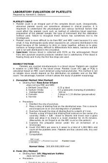
1. Disinfect the puncture site. 2. After finger puncture, wipe the 1st drop of blood. 3. Place a drop of diluent over the puncture wound. 4. The ratio of blood to diluting fluid should be 1:5. 5. Transfer the mixture into a coverslip and place it on top of a slide. 6. Allow the platelets to settle for 15-45 minutes. 30 mins: stability of diluting fluid 7. Count the RBCs and platelets under OIO until 250 RBCs have been counted.
1. Fonio’s Method Materials: Microscopic slide Pipette Diluting fluid o 14% Magnesium Sulfate Giemsa or Wright’s stain Procedure: 1. Disinfect the puncture site. 2. After finger puncture, wipe the 1st drop of blood. 3. Place a drop of diluent over the puncture wound. 4. Then press the blood the from the puncture site. 5. The ratio of blood to diluting fluid should be 1:3. 6. Make a smear using the mixture. 7. Allow the smears to dry then stain the smear. 8. Count the RBCs and platelets under OIO until 1000 RBCs have been counted. NORMAL VALUE: 250,000-500,000/ uL
2. Dameshek Method or Wet Method Materials: Microscopic slide Pipette Pipette shaker Rees Ecker fluid o Brilliant Cresyl Blue (stain) 0.1 gm o 40% Formalin 0.2 mL preservative o Sodium Citrate 100 mL To prevent clumping of platelet Procedure:
platelet
count RBC /µL = platelet counted X µL 1000
Wet mount: the RBC tend to concentrate at the edges of the cover slip if wet mount, so you have to count the RBC in the central area. Do not count on the edges so that there will be a false increase in the ratio of Platelet and RBC
3. Modified Dameshek Method
Uses siliconized medicine dropper to dilute the blood
4. Olef’s Method Materials: Microscopic slide Pipette Diluting fluid o 14% Magnesium Sulfate Giemsa or Wright’s stain Procedure: 1. Disinfect the puncture site. 2. After finger puncture, wipe the 1st drop of blood. 3. Place a drop of diluent over the puncture wound. 4. Then press the blood the from the puncture site. 5. The ratio of blood to diluting fluid should be 1:5. 6. Make a smear using the mixture. 7. Allow the smears to dry then stain the smear. 8. Count the RBCs and platelets under OIO until 1000 RBCs have been counted Same principle with Fonio's method
Confirmation of platelet count can be done on the basis of the occurrence of platelets in the peripheral smear.
25 platelet/OIO field
Thrombocytopenia Adequate Thrombocytosis
Smear estimation Materials: Microscopic slide Microscope Giemsa or Wright’s stain Procedure: 1. Make a perfect blood smear. 2. Stain with Wright stain. 3. Count platelets in 10 OIO fields.
platelet estimate= platelet counted X 2000...
Similar Free PDFs

Platelet count notes
- 6 Pages

Platelet Count hematology 2
- 2 Pages

Indirect Method Summary
- 1 Pages

The Borda Count Method
- 3 Pages

Week 6 Week 6 Week 6Week 6
- 2 Pages

Platelet Disorders - W13 ( Notes)
- 18 Pages

Week 6 ingles Week 6
- 2 Pages

Week 6 Assignment - Week 6
- 11 Pages

Tybfm SEM 6 Indirect Tax GST
- 10 Pages

Reticulocyte Count
- 2 Pages

Lecture 6 - week 6
- 32 Pages

Week 6 Study Notes - week 6
- 6 Pages

1-6 Quiz- Scientific Method
- 5 Pages
Popular Institutions
- Tinajero National High School - Annex
- Politeknik Caltex Riau
- Yokohama City University
- SGT University
- University of Al-Qadisiyah
- Divine Word College of Vigan
- Techniek College Rotterdam
- Universidade de Santiago
- Universiti Teknologi MARA Cawangan Johor Kampus Pasir Gudang
- Poltekkes Kemenkes Yogyakarta
- Baguio City National High School
- Colegio san marcos
- preparatoria uno
- Centro de Bachillerato Tecnológico Industrial y de Servicios No. 107
- Dalian Maritime University
- Quang Trung Secondary School
- Colegio Tecnológico en Informática
- Corporación Regional de Educación Superior
- Grupo CEDVA
- Dar Al Uloom University
- Centro de Estudios Preuniversitarios de la Universidad Nacional de Ingeniería
- 上智大学
- Aakash International School, Nuna Majara
- San Felipe Neri Catholic School
- Kang Chiao International School - New Taipei City
- Misamis Occidental National High School
- Institución Educativa Escuela Normal Juan Ladrilleros
- Kolehiyo ng Pantukan
- Batanes State College
- Instituto Continental
- Sekolah Menengah Kejuruan Kesehatan Kaltara (Tarakan)
- Colegio de La Inmaculada Concepcion - Cebu


