Platelet count notes PDF

| Title | Platelet count notes |
|---|---|
| Author | Trina Nogoy |
| Course | Medical Laboratory Science |
| Institution | Saint Louis University Philippines |
| Pages | 6 |
| File Size | 141 KB |
| File Type | |
| Total Downloads | 340 |
| Total Views | 437 |
Summary
LABORATORY EVALUATION OF PLATELETSPrepared by: Kenneth S. DesturaPLATELET COUNT Platelet count is an integral part of the complete blood count. Unexpectedly, abnormal platelet counts are sometimes obtained in clinical practice. It is important to understand the physiological and various technical f...
Description
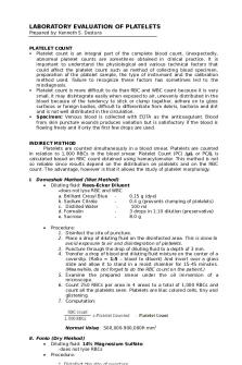
LABORATORY EVALUATION OF PLATELETS Prepared by: Kenneth S. Destura PLATELET COUNT Platelet count is an integral part of the complete blood count. Unexpectedly, abnormal platelet counts are sometimes obtained in clinical practice. It is important to understand the physiological and various technical factors that could affect the platelet count such as method of collecting blood specimen, preparation of the platelet sample, the type of instrument and the calibration method used. Failure to recognize these factors has sometimes led to the misdiagnosis. Platelet count is more difficult to do than RBC and WBC count because it is very small, it may disintegrate easily when exposed to air, unevenly distributed in the blood because of the tendency to stick or clump together, adhere on to glass surfaces or foreign bodies, difficult to differentiate from debris, bacteria and dirt and is not well distributed in the circulation. Specimen: Venous blood is collected with EDTA as the anticoagulant. Blood from skin puncture wounds produces variation but is satisfactory if the blood is flowing freely and if only the first few drops are used. INDIRECT METHOD Platelets are counted simultaneously in a blood smear. Platelets are counted in relation to 1,000 RBCs in the blood smear. Platelet Count (PC) /µL or PC/L is calculated based on RBC count obtained using hemacytometer. This method is not so reliable since results depend on the distribution on platelets and on the RBC count. The advantage, however is that it allows the study of platelet morphology. I. Dameshek Method (Wet Method) • Diluting fluid: Rees-Ecker Diluent -does not lyse RBC and WBC a. Brilliant Cresyl Blue 0.15 g (dye) b. Sodium Citrate 0.4 g (prevents clumping of platelets) c. Distilled Water 100 ml d. Formalin 3 drops in 1:10 dilution (preservative) e. Sucrose 8.0 g •
Procedure: 1. Disinfect the site of puncture. 2. Place a drop of diluting fluid on the disinfected area. This is done to avoid exposure to air and disintegration of platelets. 3. Puncture through the drop of diluting fluid to a depth of 3 mm. 4. Transfer a drop of blood and diluting fluid mixture on the center of a coverslip. (Ratio = 1:5 – blood to diluent) And invert over a glass slide and allow it to stand in a moist chamber for 15-45 minutes. (Meanwhile, do not forget to do the RBC count on the patient.) 5. Examine the prepared smear under the oil immersion of a microscope. 6. Count 250 RBCs per area in 4 areas to a total of 1,000 RBCs and count all the platelets seen. Platelets are lilac colored cells, tiny and glistening. 7. Computation:
Normal Value: 500,000-900,000/ mm3 II. Fonio (Dry Method) • Diluting fluid: 14% Magnesium Sulfate -does not lyse RBCs • Procedure:
1. Disinfect the site of puncture.
2. 3. 4. 5.
Place a drop of diluting fluid on the disinfected area. Puncture through the drop of diluting fluid to a depth of 3 mm. Transfer a drop of blood-MgSO 4 mixture on a slide. (Ratio = 1:3) Make a smear, dry and stain with Wright’s stain. (Meanwhile, do not forget to do the RBC count on the patient.) 6. Under OIO, count 250 RBCs per area in 4 areas to a total of 1,000 RBCs and count all the platelets seen. 7. Computation:
•
Normal Value: 250,000-500,000/ mm3 Advantage: 1. Easier to count RBCs and platelets 2. Size and shape of platelets can be observed
III. Olef’s Method The Olef’s method is the best method in the indirect procedures but somewhat cumbersome. Normal Value: 437,000-586,000/mm3 ESTIMATE PLATELET COUNT Ratio of Electronic Platelet Count to platelets per oil-immersion field. Before platelet estimates can be performed, the laboratory must calculate the ratio of electronic platelet count to the number of platelets per oil-immersion field of view. The procedure for calculation of this ratio and estimation factor is as follows: 1. Perform electronic platelet counts on 30 consecutive fresh patient blood samples. Make sure that the platelet count is in control. 2. For each film, under oil immersion microscopy, find an area where 50% of the red cells are overlapping in doublets or triplets. Then count the number of platelets in 10 consecutive fields. 3. Divide the total number of platelets found by 10 to obtain the average number per single oil-immersion field. 4. Divide the electronic platelet count by the average number of platelets per oil field. 5. Add the numbers obtained in step 5 and divide by 30 (the number of observations in this analysis) to obtain the average ratio of platelet count: platelets per oil-immersion field. 6. Round the number calculated to the nearest whole number to obtain the estimation factor. Estimate platelet count. Once the platelet estimation factor is calculated, the laboratory scientist may simply calculate the average number of platelets per oil-immersion field on all subsequent specimens and multiply this number by the estimation factor to obtain the platelet estimate.
Platelet Estimate of 0 – 49,000/uL 50,000 – 99,000/uL 100,000 – 149,000/uL 150,000 – 199,000/uL 200,000 – 400,000/uL 401,000 – 599,000/uL 600,000 – 800,000/uL Above 800,000/uL
Report Platelet Estimate as Marked decrease Moderate decrease Slight decrease Low normal NORMAL Slight increase Moderate increase Marked increase
DIRECT METHOD This method employs a dilution of blood using an RBC pipet with the use of a hemacytometer.
I.
Rees-Ecker Method • Diluting fluid: Rees-Ecker Diluent -does not lyse RBC and WBC a. Brilliant Cresyl Blue 0.15 g (dye) b. Sodium Citrate 0.4 g (prevents clumping of platelets) c. Distilled Water 100 ml d. Formalin 3 drops in 1:10 dilution (preservative) •
Procedure: 1. Rinse RBC pipet first with RE diluting fluid by sucking in and out the diluting fluid (to prevent disintegration of platelets). 2. Count platelets in the 25 squares in the large central square on each side of the hemocytometer. 3. Computation: Platelet Count = Platelet Counted x Dilution Factor x Area Correction Factor X 10 (Depth Correction Factor) Normal Value: 150,000-450,000/ mm3
II. Brecker Cronkite The reference method that uses phase-contrast microscopy with green (platelets appear dark) or gray filter (platelets appear pink or purple). It also uses a flat-bottomed hemacytometer whose focus can be easily adjusted and a thin coverslip (because thick coverslip retards refraction of light.). •
Diluting fluid: 1% Ammonium oxalate -lyses RBC
•
Procedure: 1. Well-mixed blood is diluted 1:100 in diluting fluid, and the vial containing the suspension is rotated on a mechanical mixer for 10 to 15 minutes. 2. The hemacytometer is filled in the usual fashion, using a separate capillary tube for each side. 3. The chamber is covered with a Petri dish for 15 minutes to allow the platelets to settle in one optical plane. A piece of wet cotton or filter paper is left beneath the dish to prevent evaporation. 4. Using this type of microscope platelets are seen very clearly against WBCs. 5. Platelets are seen as round to oval purple bodies sometimes with dendritic processes. Their internal granular structure and a purple sheen allow the platelets to be distinguished from debris, which is often refractile. Ghosts of the red cells that have been lysed by the ammonium oxalate are seen in the background. 6. Allowable difference between each chamber is +/-10 platelets. If greater than 10, repeat mixing and charging o countercheck the test. 7. Platelets are counted in 10 small squares, five on each side of the chamber. If the total number of platelets counted is less than 100, more small squares are counted until at least 100 platelets have been recorded-10 squares per side or all 25 squares in the large central square on each side of the hemocytometer, if necessary. If the total number of platelets in all 50 of these small squares is less than 50, the count should be repeated with 1:20 or 1:10 dilutions of blood. 8. Computation: Platelet Count = Platelet Counted x Dilution Factor x Area Correction Factor X 10 (Depth Correction Factor) Normal Value: 150,000-450,000/ mm3
III. Guy and Leake Method...
Similar Free PDFs

Platelet count notes
- 6 Pages

Platelet Count hematology 2
- 2 Pages

Platelet Disorders - W13 ( Notes)
- 18 Pages

Reticulocyte Count
- 2 Pages

2 - Count Zero Pdf
- 183 Pages

HEMA Reticulocyte Count
- 2 Pages

HEMA Manual Count
- 4 Pages

The Borda Count Method
- 3 Pages

Cont- Leukocyte count
- 2 Pages

Orange State - Word Count: 1837
- 4 Pages
Popular Institutions
- Tinajero National High School - Annex
- Politeknik Caltex Riau
- Yokohama City University
- SGT University
- University of Al-Qadisiyah
- Divine Word College of Vigan
- Techniek College Rotterdam
- Universidade de Santiago
- Universiti Teknologi MARA Cawangan Johor Kampus Pasir Gudang
- Poltekkes Kemenkes Yogyakarta
- Baguio City National High School
- Colegio san marcos
- preparatoria uno
- Centro de Bachillerato Tecnológico Industrial y de Servicios No. 107
- Dalian Maritime University
- Quang Trung Secondary School
- Colegio Tecnológico en Informática
- Corporación Regional de Educación Superior
- Grupo CEDVA
- Dar Al Uloom University
- Centro de Estudios Preuniversitarios de la Universidad Nacional de Ingeniería
- 上智大学
- Aakash International School, Nuna Majara
- San Felipe Neri Catholic School
- Kang Chiao International School - New Taipei City
- Misamis Occidental National High School
- Institución Educativa Escuela Normal Juan Ladrilleros
- Kolehiyo ng Pantukan
- Batanes State College
- Instituto Continental
- Sekolah Menengah Kejuruan Kesehatan Kaltara (Tarakan)
- Colegio de La Inmaculada Concepcion - Cebu


