Reticulocyte Count PDF

| Title | Reticulocyte Count |
|---|---|
| Course | Hematology 1 |
| Institution | Wichita State University |
| Pages | 2 |
| File Size | 50.3 KB |
| File Type | |
| Total Downloads | 15 |
| Total Views | 132 |
Summary
Clinical Hematology...
Description
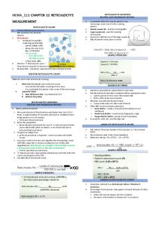
RETICULOCYTE COUNT OBJECTIVE During and upon completion of this laboratory exercise the student will: 1. 2.
Perform and interpret the results of 2 reticulocyte counts within + 10% of the value obtained by the instructor. Follow all lab safety policies as outlined in the MLS student handbook.
PRINCIPLE After the metarubricyte loses its nucleus, a small amount of RNA remains in the red cell. This cell is known as a reticulocyte. To detect the presence of this RNA, the red cells must be stained while they are still living, a process called supravital staining. After staining, the number of reticulocytes per 1,000 red cells is determined. This number is divided by ten to obtain the reticulocyte count in percent. The retic expressed as a decimal can be multiplied by the red count to obtain an absolute retic count which has more value as a diagnostic tool. REAGENTS AND EQUIPMENT 1. 2. 3. 4. 5.
Reticulocyte stain (new methylene blue) Sodium chloride 0.8 gm Potassium oxalate 1.4 gm New methylene blue N 0.5 g Distilled water 100 ml (filter before use) 6. Glass slides 7. 1 microscope 8. Small test tubes SPECIMEN Whole blood collected in EDTA, heparin or oxalate capillary blood may also be used if collected in heparinized capillary tube. PROCEDURE 1.
Place three drops of filtered retic stain in a small test tube.
2.
Add three drops of blood (may use smaller volume as long as there is an even ratio of blood to stain).
3.
Mix well and allow to stand for a minimum of 15 minutes.
4.
At the end of 15 minutes, mix the contents of the tube and make 2 slides using either a capillary tube or pasture pipets to apply the specimen.
5.
Allow smears to air dry and label correctly.
6.
Select an area where cells are evenly distributed and not overlapping. The red cells will appear as light to medium blue-green in color and the RNA present in the retics stains a deep blue. The reticulum may be abundant or sparse depending on the cells stage of development.
7.
Count all the red cells in the field which are not touching the edge. At the same time enumerate the reticulocytes on a separate counter. Continue until 500 red cells have been observed, repeat the count on another slide and if the two counts are approximately equal add them together. If not, repeat the count.
8. Calculate as follows: % reticulocyte = # of retics per 1,000 red cells 10 absolute retic = % retics as a decimal x RBC count Normal range: Adults 0.5-1.5%
Newborn infant 2.0-6.0%
DISCUSSION 1.
There are several red cell inclusion bodies that will be stained with retic stain. Howell-Jolly bodies appear as one or two round deep blue staining structures. Heinz bodies appear as light blue-green small inclusions usually at the peripheral edge of the cell. The inclusion bodies most often confused with reticulocytes are siderotic granules. If they are suspected it is best to do on Prussian blue iron stain and count the percent siderocytes and subtract that figure from your total retic percentage.
2.
Red cells are frequently noted on the reticulocyte stain which contain areas that are highly refractile. These cells should not be confused with reticulocytes. This condition is due to moisture in the air and poor drying of the smear.
3.
The range of error in reticulocyte count varies depending on the number of reticulocyte counted. There is an error of approximately + 25% on a normal retic with that range decreasing to about + 10% in a reticulocyte count of 5%.
4.
Retic stain will stain RNA, DNA, iron and denatured hgb.
Reference: McKenzie S., Clinical Laboratory Hematology, Prentice Hall, 2010, pp. 779-780....
Similar Free PDFs

Reticulocyte Count
- 2 Pages

HEMA Reticulocyte Count
- 2 Pages

Reticulocyte Production Index
- 1 Pages

2 - Count Zero Pdf
- 183 Pages

HEMA Manual Count
- 4 Pages

Platelet count notes
- 6 Pages

The Borda Count Method
- 3 Pages

Cont- Leukocyte count
- 2 Pages

Platelet Count hematology 2
- 2 Pages

Orange State - Word Count: 1837
- 4 Pages
Popular Institutions
- Tinajero National High School - Annex
- Politeknik Caltex Riau
- Yokohama City University
- SGT University
- University of Al-Qadisiyah
- Divine Word College of Vigan
- Techniek College Rotterdam
- Universidade de Santiago
- Universiti Teknologi MARA Cawangan Johor Kampus Pasir Gudang
- Poltekkes Kemenkes Yogyakarta
- Baguio City National High School
- Colegio san marcos
- preparatoria uno
- Centro de Bachillerato Tecnológico Industrial y de Servicios No. 107
- Dalian Maritime University
- Quang Trung Secondary School
- Colegio Tecnológico en Informática
- Corporación Regional de Educación Superior
- Grupo CEDVA
- Dar Al Uloom University
- Centro de Estudios Preuniversitarios de la Universidad Nacional de Ingeniería
- 上智大学
- Aakash International School, Nuna Majara
- San Felipe Neri Catholic School
- Kang Chiao International School - New Taipei City
- Misamis Occidental National High School
- Institución Educativa Escuela Normal Juan Ladrilleros
- Kolehiyo ng Pantukan
- Batanes State College
- Instituto Continental
- Sekolah Menengah Kejuruan Kesehatan Kaltara (Tarakan)
- Colegio de La Inmaculada Concepcion - Cebu


