Basic ECG - ECG PDF

| Title | Basic ECG - ECG |
|---|---|
| Course | Adult Health I |
| Institution | Chamberlain University |
| Pages | 14 |
| File Size | 755.3 KB |
| File Type | |
| Total Downloads | 96 |
| Total Views | 192 |
Summary
ECG...
Description
Electrocardiography is a science that goes back centuries. The ability to measure the electrical conduction within the heart helps health care professionals understand heart conditions and identify abnormalities quickly so they can act fast to prevent disease or death, while promoting wellness. The P wave on a rhythm strip represents the electrical stimulation of both atriums of the heart. The rhythm strip does not have any indication of muscle contraction, which is only assumed when an electrical signal is noted. The QRS wave on the rhythm strip represents the electrical stimulation of both ventricles. The T wave represents the repolarization (recharge and resting) phase of the ventricles of the heart. Repolarization of the atria occur hidden in the QRS wave. Depolarization of the atria occurs in the P wave, and depolarization of the ventricles occur in the QRS wave. A prolonged QT interval increases the risk of sudden death. The QT interval adjusts to a change in rate (like tachycardia or bradycardia). The electrocardiogram (ECG) is a graphic representation of the electricity flowing through the heart muscle. Electrocardiograms come in several different varieties which are identified by the number of leads they record, and where those leads are recorded. The most common ECG machines include an “ECG monitor” which displays 1-3 leads at a time and often includes blood pressure and other data. This is used for monitoring for rhythm changes. The other common ECG is the 12-lead electrocardiogram. This machine displays 12 views of the heart’s electrical activity at the same time and is used to diagnose more complex heart problems like myocardial infarction, or cardiomegaly. The leads are divided into two different types, limb leads, and chest leads. The limb leads attach to the extremities (or the general area of an extremity) and record the electrical activity mainly in the inferior and posterior sides of the heart muscle. The chest leads primarily record the electrical activity in the anterior and lateral sides of the heart muscle.
When looking at 3 or more leads:
It’s about TRENDS in waveform shape When looking at less than 3 leads:
It’s about rhythm and rate Rhythm strips can be recorded/viewed on electrocardiogram (ECG) monitors. The health care provider is looking for changes in heart rhythm, heart rate, or abnormal heart activity (dysrhythmias also called arrhythmias). Abnormal heart rate includes any rate that is above 100 beats per minute, called tachycardia; and any rate below 60 beats per minute, called bradycardia. Rates higher than 100 or lower than 60 are NOT ALWAYS abnormal. For example, during exercise, our rate can increase to 160 to 180 beats per minute depending on our age and physical health. Health care professionals use ECG to analyze rhythms, calculate heart rates, and in some cases, respond to changes associated with treatments. For example, a patient experiencing a myocardial infarction may have a change in the heart rhythm or rate while being monitored. In some cases, this is the first indication that there may be a change in condition and may require a more thorough nursing assessment. The electrical system of the heart starts in the sinoatrial node (SA node), a bundle of tissue that generates its own electrical signal. Once that signal travels through the atria of the heart, it reaches the atrioventricular node (AV node). This is a gateway between the top and the bottom of the heart. After a slight pause in the AV node, the electricity heads to the ventricular, down the middle (septal wall) through His bundle, and then splitting into a right and left bundles of fibers called purkinje fibers. The right bundle of fibers travel to the right side of the heart (inferior and posterior), and the left bundle of fibers travel through the left side of the heart (anterior and lateral). Problems between the sinoatrial node and the AV node can cause atrial arrhythmias.
Problems at the AV node can cause blocks. Problems after the AV node can cause ventricular arrhythmias. SA node= pacemaker How electricity flows through the heart: An electrical signal is produced in the sinoatrial node (SA node), travels through the atrium to the atrioventricular node (AV node). There it is delayed slightly to give the atrium enough time to pump (this contributes to the cardiac output). Once through the AV node, the impulse travels the bundle of HIS where it depolarizes the ventricles.
Electrocardiogram (ECG) measurement is based on milliseconds horizontally, and millimeters vertically. Milliseconds tell us how fast the electricity travels. If the electrical signal takes longer, then the tissue is not as conductive, which could be due to scar tissue, dying tissue. Some medications will also alter the speed of travel by blocking sodium and/or calcium channels in the cells. This can affect the heart rate as well.
The normal rhythm contains 3 distinct waveforms. A “P wave” which signals atrial activity (depolarization), a “QRS” wave which signals ventricular (depolarization) activity, and a “QT wave” that signals ventricular repolarization (rest). The P, QRS, and QT waves make up 1 heart cycle. The heart cycles normally between 60 and 100 times per minute, sometimes more, sometimes less.
We are most interested in three intervals: P to R interval (Beginning of the P wave to beginning of the QRS wave) QRS Interval Q to T interval (Beginning of the QRS to the end of the T wave)
What is the heart rate? There are a couple of ways to measure heart rate. Remember these as the rule of 300 and the rule of 1500. Heart rate is measured by dividing 300 by the number of BIG boxes between the "R" waves. Or you can divide 1500 by the number of SMALL boxes between the "R" waves. A lot of ECG monitors already do this for you. Note: When a rhythm is irregular, the reviewer should add several R wave measurements together to get an average.
Electrocardiogram (ECG) interpretation is a science and while there are details, a basic understanding of what is normal will help with better understanding and interpreting abnormal rhythms. Normal Sinus rhythm is: P wave for every QRS Distance between the P wave and the QRS is equal between 0.12 to 0.20ms Heart rate is between 60 and 100 beats per minute IF above is true and rate is greater than 100 it is sinus tachycardia IF above is true and rate is less than 60 it is sinus bradycardia
Think body response to environment or pathophysiology.
For Example An athlete working out could have a heart rate in the 170s, which would be a sinus tachycardia. This is normal for that athlete. A client that is sleeping may have a heart rate below 60, which is normal for sleeping. The word "sinus" indicates that the signal starts in the correct place for either condition, thereby saying that it is NOT an arrhythmia. Electrocardiogram (ECG) interpretation is a science and while there are details, a basic understanding is needed to identify high-risk changes that require immediate reporting. These include: Ventricular arrhythmias, the most common being ventricular tachycardia, always require IMMEDIATE action and are most likely to result in death. It is normal to have a ventricular tachycardia lasting 1 beat occasionally [this is called a premature ventricular contraction (PVC)]. It is when these arrhythmias are sustained (3 or more beats in a row), that symptoms can develop, and danger is present. Atrial arrhythmias, the most common being atrial fibrillation, may require urgent action. People can live their entire lives with an atrial arrhythmia, but risk factors including age and other conditions can cause complications, the worst one being a cerebral vascular accident (stroke) caused by clot formation in the left atrium from continuous atrial fibrillation over weeks, months, or years.
Heart blocks can cause significant symptoms and, in some cases, will need a device called a pacemaker to treat the client, provided other causes (including medication) are reviewed and updated. Common ECG terms: Anything with the prefix "tachy" is usually faster than 100. Anything with the prefix "brady" is usually less than 60. Ventricular always means coming from the bottom, while atrial means coming from the top. The word arrythmia usually means it is abnormal. So a ventricular arrythmias (for example) is an abnormal rhythm coming from the bottom of the heart. Ventricular tachycardia is the most common and very dangerous. Atrial fibrillation is the most common atrial arrythmia and less dangerous. Heart blocks are always about the AV node delay. Anything sinus means it's starting in the normal place. Atrial arrhythmias (the most common being atrial fibrillation) No P waves seen (hidden?), rapid or irregular (Afib) heart rate Unable to measure a PR interval (due to missing or unreadable P waves) Heart rate irregular (may be fast without visible P wave)
Think body response to environment or pathophysiology Here are some things that can cause atrial fibrillation requiring further treatment:
Pneumonia or Heart Failure Atrial arrhythmias, particularly atrial fibrillation can be caused by anything that causes atrial enlargement temporarily or chronically. In heart failure and pneumonia, fluid or infection buildup can lead to atrial dilatation and uncontrolled disorganized atrial contractions. Fever can also excite the heart.
Chronic Lung Disease Clients with lung disease tend to experience cor pulmonale, which is a high pressure in the pulmonary system (think of it as high blood pressure of the lungs). This can cause atrial irritation and enlargement leading to atrial arrhythmias and atrial fibrillation.
Medication/Drugs/Tobacco
Particularly stimulants can increase the irritability of the atria primarily by an increase in pulmonary pressures. Caffeine can be the most common and can lead to atrial fibrillation if taken in large amounts and over long periods of time. People who smoke have a 30% higher risk of atrial fibrillation. Medications like levothyroxine, used for hypothyroidism, can increase the risk of atrial fibrillation as well. Ventricular arrhythmias: No P waves seen QRS waves are wide and fast
Think dead until rhythm stabilized Here are some things that can cause life-threatening sustained ventricular arrhythmias and an example:
Electrolyte Imbalance Low magnesium levels can lead to a form or ventricular tachycardia called Torsade de Pointes (named after its unique form). This requires immediate external electricity delivered to the chest (using a defibrillator). Another electrolyte imbalance that can cause ventricular arrythmias is a low potassium level.
Altered Perfusion of the Heart Muscle Severe coronary artery disease can lead to severe decreased heart muscle perfusion, irritating the muscle causing ventricular tachycardiac – requiring immediate defibrillation.
Medication Adverse Effects or Interactions Some medications or combinations of medications prolong the QT wave making the electrical system vulnerable to a ventricular beat
which can suddenly alter the heart rhythm into ventricular tachycardia – requiring immediate defibrillation.
Genetic Predisposition There are genetic conditions that are hereditary including Long QT syndrome and hypertrophic cardiomyopathy, which can lead to ventricular arrythmias. Sometimes the first time a client knows is with a sudden death episode. This is why automatic defibrillators are found in public buildings, schools, and sports arenas. Most times there is no warning.
Electrocution High amounts of electrical amperage (lightning bolt, high voltage plug or line) can cause ventricular arrhythmias that upset the rhythm and require immediate defibrillation. Heart blocks: PR interval may or may not be the same from beat to beat Not always a QRS for every P wave QRS waves usually slow
Think slow and tired, might need a pacemaker Here are some things that can cause heart block requiring further treatment:
Myocardial Infarction Damage to the atrioventricular (AV) node caused by a myocardial infarction (usually inferior) can cause a heart block. Therefore, some clients require a pacemaker after a heart attack.
Heart Surgery Particularly aortic valve replacement surgery which causes inflammation and sometimes damage around the aortic valve near the AV node can cause heart block. All clients undergoing aortic
valve replacement have a temporary pacemaker placed in anticipation of this.
Chronic Lung Disease Along with atrial arrhythmias, chronic lung disease can cause irritation of the AV nodal area due to the high pressures in the lungs. Eventually these clients can develop sick sinus syndrome.
Medications Some medications, like beta blockers, and some calcium channel blockers, can effect the electrical activity through the AV node (bridge between the atria and ventricles). Anything affecting this bridge can lead to a partial or complete heart block. The two rhythms you never want to see! While ventricular arrythmias are not good, the two rhythms you never want to see, but may on occasion experience are ventricular fibrillation and asystole. These two rhythms require immediate call for help and starting CPR until a ‘code’ or ‘crash’ cart are available.
Tip Always check the patient’s pulse and the leads. If they have a pulse, the monitor is wrong. Disconnected leads can mimic both rhythms and checking these could save you some embarrassment. Some causes: Untreated ventricular arrythmias Same causes as ventricular arrythmias
Think HELP! Start CPR
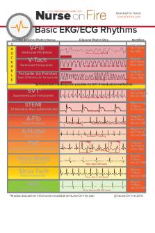
Heart block contains more P waves than QRSs, and it is slow. Ventricular fibrillation has no QRSs or P waves and is chaotic. Normal sinus rhythm has one p wave for every qrs, and the PR interval is the same. Atrial fibrillation has no P waves (the waves seen are T waves), and it is irregular. Asystole has no P waves or QRSs, and it is a flatline. Heart Block
Ventricular Fibrillation
Normal Sinus Rhythm
Atrial Fibrillation
Asystole
ST elevation means heart muscle is dying. While you may see ST elevation a single lead monitor, it is only accurate on a 12-Lead. 12lead electrocardiograms (ECG) can be challenging to read, but they do gain insight when a client has a myocardial infarction. Specifically, a 12-lead ECG will show something called ST elevation in 3 or more of the 12 leads. ST elevation is the rise of the ST segment when electricity in the heart goes around oxygen starved tissue. ST segment elevation is a sign of a myocardial infarction. This is sometimes referred to as a STEMI (ST elevation myocardial infarction). Yes, there is a non-STEMI as well which by definition is symptoms of a myocardial infarction without the ST elevation on the ECG. ST elevation in 3 or more leads on a 12-lead electrocardiogram (ECG) indicates heart cell death. When ST segments are depressed, then heart cell ischemia is occurring. This is not a normal variation, and it is not a ventricular arrythmia. Don’t treat the Electrocardiogram (ECG) rhythm, ALWAYS assess the patient FIRST After checking leads and patient pulse, start CPR for asystole and ventricular fibrillation Act urgently for atrial arrythmias Act immediately for ventricular arrhythmias
Remember that an ECG only looks at electrical signal and does not assess heart muscle or heart perfusion Remember to review medications in anyone with an ECG abnormality as some medications can cause ECG changes and abnormal rhythms...
Similar Free PDFs

Basic ECG - ECG
- 14 Pages

ECG - ECG notes
- 3 Pages

Basic EKG ECG Rhythms Cheatsheet
- 1 Pages

ECG 1 - Atividades ECG- cardiologia
- 12 Pages

ECG 2020 - ECG - LE TOURNEAU
- 7 Pages

Informe ECG
- 22 Pages

ECG - electrocardiograma
- 5 Pages

ECG pléthysmographie
- 2 Pages

ECG taller
- 11 Pages

Cardiologia - ECG
- 59 Pages

ECG simulation
- 13 Pages

Electrocardiograma (ECG)
- 4 Pages

ECG PDF Edition
- 59 Pages
Popular Institutions
- Tinajero National High School - Annex
- Politeknik Caltex Riau
- Yokohama City University
- SGT University
- University of Al-Qadisiyah
- Divine Word College of Vigan
- Techniek College Rotterdam
- Universidade de Santiago
- Universiti Teknologi MARA Cawangan Johor Kampus Pasir Gudang
- Poltekkes Kemenkes Yogyakarta
- Baguio City National High School
- Colegio san marcos
- preparatoria uno
- Centro de Bachillerato Tecnológico Industrial y de Servicios No. 107
- Dalian Maritime University
- Quang Trung Secondary School
- Colegio Tecnológico en Informática
- Corporación Regional de Educación Superior
- Grupo CEDVA
- Dar Al Uloom University
- Centro de Estudios Preuniversitarios de la Universidad Nacional de Ingeniería
- 上智大学
- Aakash International School, Nuna Majara
- San Felipe Neri Catholic School
- Kang Chiao International School - New Taipei City
- Misamis Occidental National High School
- Institución Educativa Escuela Normal Juan Ladrilleros
- Kolehiyo ng Pantukan
- Batanes State College
- Instituto Continental
- Sekolah Menengah Kejuruan Kesehatan Kaltara (Tarakan)
- Colegio de La Inmaculada Concepcion - Cebu


