Atomic Absorption Spectroscopy PDF

| Title | Atomic Absorption Spectroscopy |
|---|---|
| Author | Dr. Kalsoom Saleem |
| Pages | 9 |
| File Size | 793.5 KB |
| File Type | |
| Total Downloads | 3 |
| Total Views | 447 |
Summary
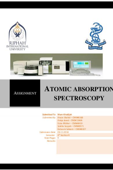
ATOMIC ABSORPTION SPECTROSCOPY (AAS) ASSIGNMENT ATOMIC ABSORPTION SPECTROSCOPY v, 2014 Submitted To Mam Khadijah Submitted By Anum Shahid – CMS#8168 Aniqa Javed - CMS#13464 Faiza Iftikhar – CMS#8420 Saleha Sayyab – CMS#8571 Kalsoom Saleem – CMS#8107 Submission Date 06-11-2014 Semester 6th Section A ...
Description
ATOMIC ABSORPTION SPECTROSCOPY (AAS)
ASSIGNMENT
ATOMIC ABSORPTION SPECTROSCOPY
v, 2014 Submitted To Mam Khadijah Submitted By Anum Shahid – CMS#8168 Aniqa Javed - CMS#13464 Faiza Iftikhar – CMS#8420 Saleha Sayyab – CMS#8571 Kalsoom Saleem – CMS#8107 Submission Date 06-11-2014 Semester 6th Section A Total Pages 9 Remarks
1|Page
ATOMIC ABSORPTION SPECTROSCOPY (AAS)
Table of Contents Sr #
Content
Page #
1
Introduction
3
2
Basic Principle of AAS
4
3
Atomic Spectra
4
4
Instrumentation of AAS
5
5
Interpretation of AAS
7
6
Applications of AAS
8
7
References
9
2|Page
ATOMIC ABSORPTION SPECTROSCOPY (AAS) 1. Introduction Atomic-absorption spectroscopy quantifies the absorption of ground state atoms in the gaseous state. The atoms absorb ultraviolet or visible light and make transitions to higher electronic energy levels. The analyte concentration is determined from the amount of absorption. Concentration measurements are usually determined from a working curve after calibrating the instrument with standards of known concentration. Atomic absorption is a very common technique for detecting metals and metalloids in environmental samples. When a solution containing metallic species are introduced into a flame, the vapor of metallic species will be obtained. Some of the metal atoms may be raised to higher energy level and emit characteristic radiation. However, large amount of metal atoms will remain in non-emitting ground state. These ground state atoms of particular element are receptive of light radiation of their own specific resonance wavelength. Thus, when a light of this wavelength passed through a flame having atom of metallic species, part of light will be absorbed and the absorption will be proportional to the density of atom in the flame.
Elements determined from this technique as shown in Table 1.
Aluminium (Al) Antimony (Sb)
Elements that can be detected by AAS Copper (Cu) Mercury (Hg) Cadmium (Cd) Gallium (Ga) Molybdenum (Mo) Cobalt (Co)
Calcium (Ca) Chromium (Cr)
Arsenic (As)
Hafnium (Hf)
Niobium (Nb)
Nickel (Ni)
Lead (Pb)
Beryllium (Be)
Indium (In)
Ruthenium (Ru)
Manganese (Mn)
Lithium (Li)
Barium (Ba)
Iron (Fe)
Tin (Sn)
Magnesium (Mg)
Vanadium (V)
Tungsten (W)
Vanadium (V)
Zinc (Zn)
Zirconium (Zr) 3|Page
ATOMIC ABSORPTION SPECTROSCOPY (AAS) The atoms in the atomizer get promoted to higher orbitals (excited state) for a short period of time (nanoseconds) by absorbing a defined quantity of energy (radiation of a given wavelength). This amount of energy, i.e., wavelength, is specific to a particular electron transition in a particular element. In general, each wavelength corresponds to only one element, and the width of an absorption line is only of the order of a few Pico meters (pm), which gives the technique its elemental selectivity. The radiation flux without a sample and with a sample in the atomizer is measured using a detector, and the ratio between the two values (the absorbance) is converted to analyte concentration or mass using the Beer-Lambert Law. 2. Principle The Beer–Lambert law: Atomic absorption spectroscopy relies on the Beer-Lambert law to determine the concentration of a particular analyte in a sample. The absorption spectrum and molar absorbance of the desired sample element are known, a known amount of energy is passed through the atomized sample and by then measuring the quantity of light, it is possible to determine the concentration of the element being measured. There is a linear relationship between absorbance and concentration of an absorbing species.
A= Absorbance l= path lenth of cell (cm) c=molar concentration = wavelength-dependent molar absorptivity coefficient
Applying Lambert-Beer’s law in atomic absorption spectroscopy is difficult due to variations in the atomization from the sample matrix and non-uniformity of concentration and path length of analyte atoms. Concentration measurements are usually determined from a calibration curve generated with standards of known concentration. 3. Atomic spectra Atomic spectra feature sharp bands. For example hydrogen spectrum: n=1 energy DE
n=2 n=3
4|Page
ATOMIC ABSORPTION SPECTROSCOPY (AAS) 4. Instrumentation of AAS Atomic absorption instruments consist of a a. Radiation Source b. Monochromator c. Flame or electrothermal atomizer in which sample is introduced d. Atomizer e. Detector a. Radiation Source Although radiation in the UV-Vis region is required, we cannot use broadband sources. This is because even the best monochromators cannot provide a bandwidth that is narrower than the atomic absorption line. If the bandwidth of the incident radiation is wider than the line width, measurement will fail as absorption will be only a tiny fraction of a large signal which is difficult to measure and will result in very low sensitivities (figure a). Therefore, line sources with bandwidths narrower than that of the absorption lines must be used (figure b). This can be achieved by using a lamp producing the emission line of the element of interest where analyte atoms can absorb that line. Conditions are established to get a narrower emission line than the absorption line. This can in fact be achieved by getting an emission line of interest at the following conditions: 1. Low temperatures: to decrease Doppler broadening (which is easily achievable since the temperature of the source is always much less than the temperature in flames). 2. Lower pressures: this will decrease pressure broadening and will thus produce a very narrow emission line. Atomic Line Width Monochromator Bandwidth (different Scales) this may suggest the need for a separate lamp for each element, which is troublesome and inconvenient. However, recent developments lead to introduction of multi-element lamps. In this case, the lines from all elements should not interfere and must be easily resolved by the monochromator so that, at a specific time, a single line of one element is leaving the exit slit. Hollow Cathode Lamp (HCL) This is the most common source in atomic absorption spectroscopy. It is formed from a tungsten anode and a cylindrical cathode the interior surface of which is coated by the metal of interest. The two electrodes are usually sealed in a glass tube with a quartz window and filled with argon at low pressure (1-5 torr). Ionization of the argon is forced by application of 5|Page
ATOMIC ABSORPTION SPECTROSCOPY (AAS) about 300 V DC where positively charged Ar+ heads rapidly towards the negatively charged cathode causing sputtering. A portion of sputtered atoms is excited and thus emits photons as atoms relax to ground state. The cylindrical shape of the cathode serves to concentrate the beam in a limited region and enhances redisposition of sputtered atoms at the hollow surface. High potentials usually result in high currents, which, in turn, produce more intense radiation. However, Doppler broadening increases as a result. In addition, the higher currents will produce high proportion of unexcited atoms that will absorb some of the emission beam, which are referred to as selfabsorption (a lower intensity at the center of the line is observed in this case). b. Monochromators This is a very important part in an AA spectrometer. It is used to separate out all of the thousands of lines. Without a good monochromator, detection limits are severely compromised. A monochromator is used to select the specific wavelength of light, which is absorbed by the sample, and to exclude other wavelengths. The selection of the specific light allows the determination of the selected element in the presence of others. c. Atomizer Atomization is separation of particles into individual molecules and breaking molecules into atoms.This is done by exposing the analyte to high temperatures in a flame or graphite furnace. The role of the atom cell is to primarily dissolvate a liquid sample and then the solid particles are vaporized into their free gaseous ground state form. In this form, atoms will be available to absorb radiation emitted from the light source and thus generate a measurable signal proportional to concentration. There are two types of atomization: Flame and Graphite furnace atomization. d. Flame Or Electrothermal Atomizer In Which Sample Is Introduced There can be significant amounts of emission produced in flames due to presence of flame constituents (molecular combustible products) and sometimes impurities in the burner head. This emitted radiation must be removed for successful sensitive determinations by AAS, otherwise a negative error will always be observed. The detector will see the overall signal, which is the power of the transmitted beam (P) in addition to the power of the emitted radiation from flame (Pe). Therefore if we are measuring absorbance, this will result in a negative error as the detector will measure what it appears as a high transmittance signal (actually it is P + Pe). In case of emission measurements, there will always be a positive
6|Page
ATOMIC ABSORPTION SPECTROSCOPY (AAS) error since emission from flame is an additive value to the actual sample emission. It is therefore obvious that we should get rid of this interference from emission in flames. e. Detector The light selected by the monochromator is directed onto a detector that is typically a photomultiplier tube, whose function is to convert the light signal into an electrical signal proportional to the light intensity. A signal amplifier fulfills the processing of electrical signal. The signal could be displayed for readout, or further fed into a data station for printout by the requested format.
5. Interpretation of AAS Atomic theory tells us that the electrons in all atoms are in well-defined orbitals. For example, in uranium, the electron shells with principal quantum number 1-6 are all filled and the shell with principal quantum number 7 is partially filled. Numerous orbital are available in each shell that are s, p, d orbitals, etc. in the filled shell, each orbital accommodates an electron. In the unexcited atoms, these electrons reside in the orbital with the lowest energy level. However, the upper empty orbital is available to accommodate an electron. During excitation the electrons with the lowest electron moves from normal low-energy level to an orbital with a higher energy. This orbital may be in the same shell or in a higher shell, inasmuch as each orbital is available to accommodate an electron unless excluded by quantum theory-forbidden transitions. Example: In atomic sodium, electron fill the shells with quantum numbers 1 and 2, and one electron is in the shell with the quantum number 3. When the sodium is in the ground state, this will be in orbital with the lowest energy, i.e., 3s. if we excite sodium, the 3s electron can move to n orbital with higher energy. The energy level next to the 3s level is the 3p energy level, hence it is possible for an electron to go from a 3s to 3p orbital. It is also possible for the 3s electron to go into orbitals of even higher 7|Page
ATOMIC ABSORPTION SPECTROSCOPY (AAS) energy, such as 4p, 4d, 5p, 5d, etc. When the valence electrons of sodium is in the 3s orbital, its lowest energy state, the sodium is said to be in the ground state. When the electron is in any orbital with higher energy, the sodium is said to be in excited energy state, or excited sodium. When radiation energy is absorbed, the atom becomes excited. If we use a prism to disperse the radiation falling on the atoms, the absorption spectrum appears as a series of narrow lines opposed to wide bands. If the transition is between the ground state and lowest excited state, then it is said that the absorption line is the resonance line. Transitions between the ground state and the upper excited states are possible but are not often used. The energy levels of sodium are shown in figure, for sake of clarity, upper state transitions are omitted. In sodium, the transition between the 3s orbital and a 3p orbital can be achieved by absorbing radiations at 589 or 589.5 nm. Similar absorption of radiation at 33.03 nm will cause sodium to be excited from the 3s ground state to the 5p excited state orbital. Transitions between the 3s orbital and orbitals with principal quantum number 6 requires more energy. 6. Applications 1. It is used for water analysis for the presence of following content (e.g. Ca, Mg, Fe, Si, Al, Ba ) 2. Also used in food analysis and soil analysis 3. In clinical analysis it is used for analyzing metals in biological fluids and tissues such as whole blood, plasma, urine, saliva, brain tissue, liver, muscle tissue, semen 4. In Pharmaceuticals it has applications in some pharmaceutical manufacturing processes, minute quantities of a catalyst that remain in the final drug product 5. It is used in petroleum industry, metallic impurities in petrol, lubricating oils have been determined. 6. It is used in alloys, metallurgy and in inorganic analysis. 7. Used in analysis of many ores and minerals. 8. It is used in biochemical analysis such as used in estimation of sodium, potassium, zinc, lead, cadmium, mercury, calcium, iron and magnesium. 9. Also used in pharmaceutical analysis, for estimation of zinc in insulin preparations, creams and in calamine, oils, calcium in calcium salts, lead in calcium carbonate and also as impurity in number of chemical salts have been done. 10. Sodium, calcium, and potassium in saline and ringer solutions are estimated by this method. 11. Analysis of ash for determining the contents of sodium, potassium, calcium, magnesium and iron is carried out in boiler deposits. 12. Used in cement industry. 13. Used in agriculture, soil, forestry, fertilizer and oceanography etc. 14. Used in assay of intraperitoneal dialysis, activated charcoal, cisplatin. 8|Page
ATOMIC ABSORPTION SPECTROSCOPY (AAS) 7. References a. James W. Robinson, Atomic Spectroscopy, Second Edition, page 100-102 b. M.Arora, Aseem Anand, Instrumental Method Of Chemical Analysis, Himalaya Publishin House, Fifth Edition, 2005, Page 240-242 c. Aurora Biomed Inc., Aurora Instruments. 2014. http://www.aurorabiomed.com/atomicabsorption-spectroscopy/ d. Hitachi High-Technologies Corporation. 2011-2014. http://www.hitachi-hitec.com/global/science/aas/aas_basic_3.html e. Galbraith Laboratories, Inc. 2011-2014. http://www.galbraith.com/spectroscopy.htm f. M.Arora, Aseem Anand, Instrumental Method Of Chemical Analysis, Himalaya Publishin House, Fifth Edition, 2005, Page 240-241
9|Page...
Similar Free PDFs

Atomic Absorption Spectroscopy
- 271 Pages

Atomic Absorption Spectroscopy
- 9 Pages

Atomic Absorption Spectroscopy
- 1 Pages

Chem 1411 Atomic Spectroscopy
- 3 Pages

Spectroscopy
- 5 Pages

Mass Spectroscopy
- 14 Pages

Cour absorption
- 15 Pages

TD Absorption
- 4 Pages

Spectroscopy Lab
- 2 Pages
Popular Institutions
- Tinajero National High School - Annex
- Politeknik Caltex Riau
- Yokohama City University
- SGT University
- University of Al-Qadisiyah
- Divine Word College of Vigan
- Techniek College Rotterdam
- Universidade de Santiago
- Universiti Teknologi MARA Cawangan Johor Kampus Pasir Gudang
- Poltekkes Kemenkes Yogyakarta
- Baguio City National High School
- Colegio san marcos
- preparatoria uno
- Centro de Bachillerato Tecnológico Industrial y de Servicios No. 107
- Dalian Maritime University
- Quang Trung Secondary School
- Colegio Tecnológico en Informática
- Corporación Regional de Educación Superior
- Grupo CEDVA
- Dar Al Uloom University
- Centro de Estudios Preuniversitarios de la Universidad Nacional de Ingeniería
- 上智大学
- Aakash International School, Nuna Majara
- San Felipe Neri Catholic School
- Kang Chiao International School - New Taipei City
- Misamis Occidental National High School
- Institución Educativa Escuela Normal Juan Ladrilleros
- Kolehiyo ng Pantukan
- Batanes State College
- Instituto Continental
- Sekolah Menengah Kejuruan Kesehatan Kaltara (Tarakan)
- Colegio de La Inmaculada Concepcion - Cebu


